. 2017 Dec 28;10(1):625.
doi: 10.1186/s13071-017-2547-0.

Acanthamoeba-mediated cytopathic effect correlates with MBP and AhLBP mRNA expression

Affiliations

Acanthamoeba-mediated cytopathic effect correlates with MBP and AhLBP mRNA expression

Sook-Luan Ng et al. Parasit Vectors. .

Abstract

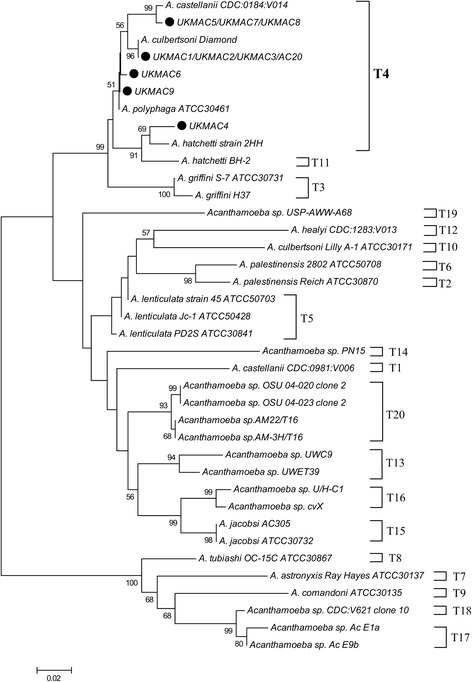
Background: In recent years, the concern of Acanthamoeba keratitis has increased since the infection is often associated with contact lens use. Partial 18S rRNA genotypic identification of Acanthamoeba isolates is important to correlate with pathophysiological properties in order to evaluate the degree of virulence. This is the first report of genotypic identification for clinical isolates of Acanthamoeba from corneal scrapings of keratitis in Malaysia. This study is also the first to correlate the mRNA expression of MBP and AhLBP as virulent markers for axenic strains of Acanthamoeba.

Results: In this study, ten clinical isolates were obtained from corneal scrapings. Rns genotype and intra-genotypic variation at the DF3 region of the isolates were identified. Results revealed that all clinical isolates belonged to the T4 genotype, with T4/6 (4 isolates), T4/2 (3 isolates), T4/16 (2 isolates) and one new genotype T4 sequence (T4/36), being determined. The axenic clinical isolates were cytopathogenic to rabbit corneal fibroblasts. MBP and AhLBP mRNA expression are directly correlated to Acanthamoeba cytopathic effect.

Conclusions: All ten Malaysian clinical isolates were identified as genotype T4 which is predominantly associated with AK. Measuring the mRNA expression of Acanthamoeba virulent markers could be useful in the understanding of the pathogenesis of Acanthamoeba keratitis.

Keywords: Acanthamoeba; AhLBP; Cytopathic; Genotype; Keratitis; MBP.


Conflict of interest statement

Ethics approval

This project was approved by the Ethics and Research Committee of Universiti Kebangsaan Malaysia Medical Centre, reference number FF-274-2010. The use of rabbit corneal cells was approved by UKM Animal Ethics Committee with reference number FP/FISIO/2012/CHUA/18-JANURAY/420-JANUARY-2012-JUNE-2013-AR-CAT2.

Consent for publication

Not applicable.

Competing interests

The authors declare that they have no competing interests.

Publisher’s Note

Springer Nature remains neutral with regard to jurisdictional claims in published maps and institutional affiliations.

Figures

Fig. 1
Fig. 1
Evolutionary relationships of taxa. The evolutionary history was inferred using the neighbor-joining method for the 18S rDNA partial sequences of clinical isolates (black dots). The optimal tree with the sum of branch length = 1.25072993 is shown. The percentage of replicate trees in which the associated taxa clustered together in the bootstrap test (1000 replicates) is shown next to the branches. The tree is drawn to scale, with branch lengths in the same units as those of the evolutionary distances used to infer the phylogenetic tree. The evolutionary distances were computed using the Kimura 2-parameter method and are in the units of the number of base substitutions per site. The analysis involved 38 nucleotide sequences. There were a total of 309 positions in the final dataset. Evolutionary analyses were conducted with MEGA7
Fig. 2
Fig. 2
T4 subgenotype of clinical isolates. Primary sequence alignment of a subset of the highly variable and informative region of DF3 (stem 29-1, 18S rRNA) of Malaysian clinical isolates from corneal scrapings and the reference sequences of the T4 Rns genotype. Abbreviations for samples and reference sequences are as defined in Table 1. Sequences were aligned by similarity. Asterisks denote similar positions and gaps are represented as dashes. Samples of the present study are indicated in bold
Fig. 3
Fig. 3
Morphology of cysts and axenic trophozoites of group II and III Acanthamoeba stained with methylene blue and observed under phase contrast microscopy (magnification ×1000). a Thick ectocyst and polygonal endocyst in Group II. b Thin ectocyst adjacent to the endocyst in Group III. c, d Acanthamoeba trophozoites with characteristic acanthapodia, contractile vacuole and prominent nucleus. Scale-bars: 20 μm
Fig. 4
Fig. 4
Cytopathic effect of Acanthamoeba trophozoites on corneal fibroblasts. The seeding density of Acanthamoeba was 105 (a) and 106 trophozoites (b) for 3, 6 and 24 h co-culture. AC20 had a significantly higher CPE than all other isolates (*P < 0.05)
Fig. 5
Fig. 5
Cytopathic effect of AC20 isolate on the corneal fibroblasts with seeding of 106 trophozoites. a The corneal fibroblasts are fusiform; the Acanthamoeba trophozoites with prominent contractile vacuoles were feeding on the edge of cells and thus forming multiple small lesions on the monolayer cells after 3 h co-culture. b The gaps between keratocytes were increased with the time due to the cytopathic effect of trophozoites after 6 h co-culture. c All corneal fibroblasts were lysed by trophozoites after 24 h co-culture and only left trophozoites in the vessel. Giemsa staining (magnification ×100). Scale-bars: 200 μm
Fig. 6
Fig. 6
Cytopathic effect (a), (b) growth rate, mRNA expression of MBP (c) and AhLBP (d) virulent markers of clinical isolates at 24 h. AC20 had a statistically significant higher CPE, growth rate and MBP expression as compared to the other isolates (* P < 0.05). UKMAC4 had a statistically significant lower growth rate and MBP expression as compared to the other isolates (# P < 0.05)
Fig. 7
Fig. 7
Agrose gel electrophoresis of PCR products. The products were for the target gene ASA.1 for ten Acanthamoeba isolates (a) and the housekeeping gene ARP2, Acanthamoeba virulent markers MBP and AhLBP genes (b). A single band of PCR product indicated the specificity of the synthesised primers

Similar articles

Cited by

References

    1. Lorenzo-Morales J, Khan NA, Walochnik J. An update on Acanthamoeba keratitis: diagnosis, pathogenesis and treatment. Parasite. 2015;22:10. doi: 10.1051/parasite/2015010. - DOI - PMC - PubMed
    1. Anisah N, Yusof S, Rahimah I, Norhayati M. Isolation of Acanthamoeba spp. from domestic water tap. Trop Biomed. 2003;20(1):87–89.
    1. Khan NA. Pathogenesis of Acanthamoeba infections. Microb Pathog. 2003;34(6):277–285. doi: 10.1016/S0882-4010(03)00061-5. - DOI - PubMed
    1. Illingworth CD, Cook SD. Acanthamoeba keratitis. Surv Ophthalmol. 1998;42(6):493–508. doi: 10.1016/S0039-6257(98)00004-6. - DOI - PubMed
    1. Seal D. Acanthamoeba keratitis update - incidence, molecular epidemiology and new drugs for treatment. Eye. 2003;17(8):893–905. doi: 10.1038/sj.eye.6700563. - DOI - PubMed

Publication types

MeSH terms

LinkOut - more resources