Review
. 2018 Feb;19(2):206-224.
doi: 10.15252/embr.201745302. Epub 2017 Dec 27.

The bright and the dark side of human antibody responses to flaviviruses: lessons for vaccine design

Affiliations
Review

The bright and the dark side of human antibody responses to flaviviruses: lessons for vaccine design

Félix A Rey et al. EMBO Rep. 2018 Feb.

Abstract

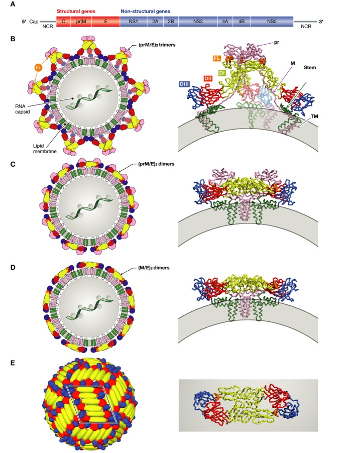
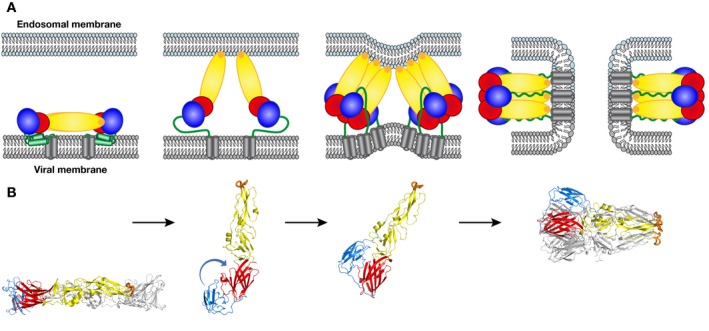
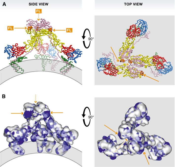
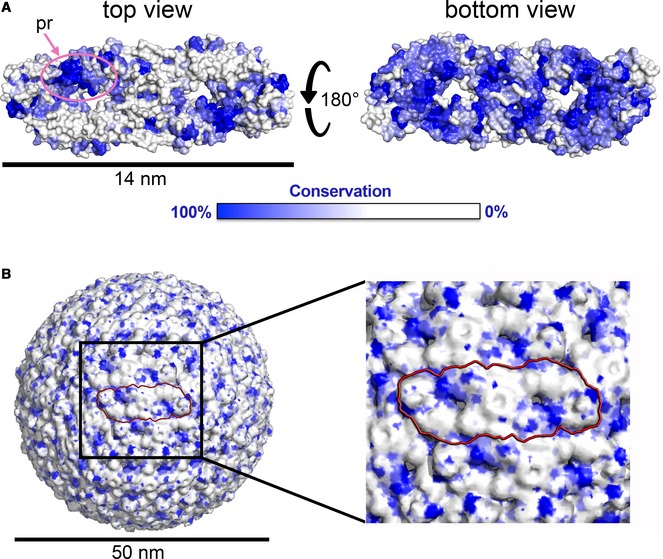
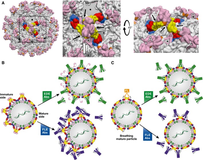
Zika and dengue viruses belong to the Flavivirus genus, a close group of antigenically related viruses that cause significant arthropod-transmitted diseases throughout the globe. Although infection by a given flavivirus is thought to confer lifelong protection, some of the patient's antibodies cross-react with other flaviviruses without cross-neutralizing. The original antigenic sin phenomenon may amplify such antibodies upon subsequent heterologous flavivirus infection, potentially aggravating disease by antibody-dependent enhancement (ADE). The most striking example is provided by the four different dengue viruses, where infection by one serotype appears to predispose to more severe disease upon infection by a second one. A similar effect was postulated for sequential infections with Zika and dengue viruses. In this review, we analyze the molecular determinants of the dual antibody response to flavivirus infection or vaccination in humans. We highlight the role of conserved partially cryptic epitopes giving rise to cross-reacting and poorly neutralizing, ADE-prone antibodies. We end by proposing a strategy for developing an epitope-focused vaccine approach to avoid eliciting undesirable antibodies while focusing the immune system on producing protective antibodies only.

Keywords: antibody neutralization; antibody‐dependent enhancement; flavivirus structure; particle heterogeneity; vaccine design.


Figures

Figure 1
Figure 1. Flavivirus particle assembly
(A) The flavivirus open‐reading frame coding for a single precursor polyprotein. Co‐ and post‐translational proteolytic processing results in the various proteins indicated, with structural and non‐structural proteins in red and blue, respectively. (B–E) Sketches representing the flavivirus particle at different maturation stages (left), with a ribbon representation of the relevant envelope protein complexes on the right. The genomic ribonucleoprotein complex with protein C has not been visualized and is represented here within a central gray circle (in B–D), although its organization is unknown. Protein E is colored according to domains: red, yellow, blue, and green for domains I, II, III, and stem/TM (transmembrane anchor), respectively. The fusion loop is highlighted in orange, and prM/M (including its TM region) is shown in pink. The viral membrane is represented in gray. (B) Left: The immature flavivirus particle as it buds in the ER of the infected cells. Right: A single (prM/E)3 spike is displayed as ribbons (PDB code 4B03). (C) Left: The immature flavivirus particle after exposure to the acidic pH of the trans‐Golgi apparatus, where the trimeric spikes dissociate and the 180 prM/E heterodimers re‐associate into 90 (prM/E)2 dimers. Right: A single (prM/E)2 dimer is shown as ribbons (PDB codes 3C6R and 3JP2). (D) Left: The mature flavivirus particle with 90 (M/E)2 dimers. Right: A single (M/E) 2 dimer is shown in ribbons. (E) Left: Herringbone pattern of E dimers on the surface of mature virus particles, consisting of 30 rafts of three E dimers. One raft is framed in black. Right: Top view of a single E homodimer shown as ribbons.
Figure 2
Figure 2. The fusogenic conformational change of the E protein during cell entry
The E protein is colored as in Fig 1. (A) Schematic of the fusion process: A mature E dimer anchored in the viral membrane is represented in the left panel. The dimer dissociates upon exposure to acidic pH in the endosome, inserting the fusion loop into the endosomal membrane (second panel). The aligned E monomers then trimerize, thereby creating a binding site for domain III at the sides of a “core trimer”. Domain III then flips to the sides of the trimer, pulling the stem and TM segments toward the endosomal membrane (third panel). The final, post‐fusion conformation, brings the viral TM segment next to the fusion loop, inducing first hemi‐fusion (i.e., fusion of only the outer leaflets of the two membranes) followed by opening of a fusion pore (fourth panel). The final post‐fusion conformation of E is achieved only after fusion pore formation. (B) 3D structures of the dengue virus 2 E ectodomains (lacking the stem/TM regions) matching the steps indicated in (A) (PDB codes 1OAN and 1OK8 for pre‐fusion and post‐fusion conformations, respectively).
Figure 3
Figure 3. The fusion loop is exposed in the immature (prM/E)3 spikes
Left panels: side views, right panels: top views. (A) The spike shown in ribbons as in Fig 1. (B) The spike shown in a surface representation colored according to amino acid conservation across all flaviviruses, from white (variable) to dark blue (absolutely conserved). Patches corresponding to the fusion loop are indicated by orange arrows. One of the three fusion loops in the trimer is not exposed (orange dashed arrow).
Figure 4
Figure 4. The mature particle exposes a conserved patch
(A) The E dimer in surface representation colored according to amino acid conservation across all flaviviruses, in its top and bottom view. The pr binding site is encircled in pink. This site corresponds to the conserved E dimer epitope (EDE) discussed in the text. Notice the high conservation of the E dimer “underside” in the “bottom” view (right panel), which faces the viral membrane in the particle. (B) The mature flavivirus particle shown in surface representation, colored as in (A). One dimer is highlighted by a red contour. The black box indicates the region zoomed in the right panel.
Figure 5
Figure 5. Differential binding modes of EDE versus FLE antibodies (Abs)
(A) Left panel: Intertwined lattice of (prM/E)3 trimers in the spiky immature particles. A central (prM/E)3 spike is represented with protein E colored by domains as in Fig 1. All other spikes have protein E in gray. prM is displayed in pink throughout. A box highlights the region zoomed in the middle panel. Middle panel: intertwined array of (prM/E)3 spikes, in which the three adjacent trimers (A, B, and C) interact underneath the central one (black arrows), underpinning it. In partially immature spikes, such stabilization is lost for the trimers at the interface between spiky and mature patches. Right panel: View along the white arrow of the middle panel, rotated by 45°. The black arrows point to the contacts between adjacent spikes. (B) Interaction of partially mature particles with EDE (green) and FLE (blue) antibodies. The spikes at the interface between mature and immature patches do not have the underpinning and stabilizing effect of three surrounding spikes shown in (A), and are therefore destabilized at these edge regions. The very high affinity of the EDE antibodies for the E dimer appears to shift the trimer‐dimer equilibrium toward dimers at the interface. Inner trimers become destabilized in turn upon EDE antibody binding, resulting in a domino effect such that the particle ends up fully coated with antibodies, as illustrated in the middle, top panel. FLE antibodies, in contrast, can readily bind to the immature patches, but also to the mature side depending on the extent of breathing (see panel C). (C) Interaction of mature particles with EDE (green) and FLE (blue) antibodies. Particle breathing (indicated by curved black arrows) leads to transient exposure of the fusion loop. EDE antibodies bind readily, regardless of the breathing behavior of the E dimers. FLE antibodies can only bind when the fusion loop is exposed through breathing, allowing particle internalization via Fcγ receptor‐mediated endocytosis before attaining sufficient antibody coating for neutralization depending on the breathing kinetics, giving rise to ADE. This situation can also explain why neutralizing antibodies, if present with ADE‐prone antibodies such as the FLEs, can override ADE by completing the antibody coating, without the need to displace the bound FLE antibody.

References

    1. Bhatt S, Gething PW, Brady OJ, Messina JP, Farlow AW, Moyes CL, Drake JM, Brownstein JS, Hoen AG, Sankoh O et al (2013) The global distribution and burden of dengue. Nature 496: 504–507 - PMC - PubMed
    1. Weaver SC, Costa F, Garcia‐Blanco MA, Ko AI, Ribeiro GS, Saade G, Shi P‐Y, Vasilakis N (2016) Zika virus: history, emergence, biology, and prospects for control. Antiviral Res 130: 69–80 - PMC - PubMed
    1. WHO (2017) Yellow fever in Africa and the Americas, 2016. Wkly Epidemiol Rec 92: 442–452 - PubMed
    1. ECDC (2017) Rapid risk assessment: outbreak of yellow fever in Brazil. First update, 13 April 2017. Stockholm: ECDC;
    1. Wilder‐Smith A, Gubler DJ, Weaver SC, Monath TP, Heymann DL, Scott TW (2017) Epidemic arboviral diseases: priorities for research and public health. Lancet Infect Dis 17: e101–e106 - PubMed

Publication types

MeSH terms