. 2018 Feb 9;293(6):2159-2171.
doi: 10.1074/jbc.M117.808881. Epub 2017 Dec 27.

Arabidopsis thaliana rapid alkalinization factor 1-mediated root growth inhibition is dependent on calmodulin-like protein 38

Affiliations

Arabidopsis thaliana rapid alkalinization factor 1-mediated root growth inhibition is dependent on calmodulin-like protein 38

Wellington F Campos et al. J Biol Chem. .

Abstract

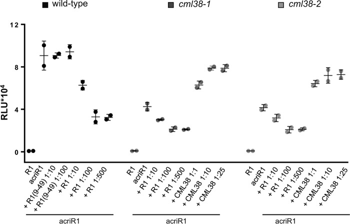
Arabidopsis thaliana rapid alkalinization factor 1 (AtRALF1) is a small secreted peptide hormone that inhibits root growth by repressing cell expansion. Although it is known that AtRALF1 binds the plasma membrane receptor FERONIA and conveys its signals via phosphorylation, the AtRALF1 signaling pathway is largely unknown. Here, using a yeast two-hybrid system to search for AtRALF1-interacting proteins in Arabidopsis, we identified calmodulin-like protein 38 (CML38) as an AtRALF1-interacting partner. We also found that CML38 and AtRALF1 are both secreted proteins that physically interact in a Ca2+- and pH-dependent manner. CML38-knockout mutants generated via T-DNA insertion were insensitive to AtRALF1, and simultaneous treatment with both AtRALF1 and CML38 proteins restored sensitivity in these mutants. Hybrid plants lacking CML38 and having high accumulation of the AtRALF1 peptide did not exhibit the characteristic short-root phenotype caused by AtRALF1 overexpression. Although CML38 was essential for AtRALF1-mediated root inhibition, it appeared not to have an effect on the AtRALF1-induced alkalinization response. Moreover, acridinium-labeling of AtRALF1 indicated that the binding of AtRALF1 to intact roots is CML38-dependent. In summary, we describe a new component of the AtRALF1 response pathway. The new component is a calmodulin-like protein that binds AtRALF1, is essential for root growth inhibition, and has no role in AtRALF1 alkalinization.

Keywords: calmodulin (CaM); cell growth; cell signaling; development; peptide hormone; rapid alkalinization factor; root; secreted protein; secretory pathway.


Conflict of interest statement

The authors declare that they have no conflicts of interest with the contents of this article

Figures

Figure 1.
Figure 1.
The AtRALF1 peptide interacts with the CML38 protein. A and B, yeast two-hybrid assay on complete (+His) or selective medium (−His). AD, activation domain construct; BD, binding domain construct. C, pulldown assay with the recombinant proteins AtRALF1 and GST-CML38 in the presence of Ca2+ or the Ca2+ chelator EGTA. D, pulldown assay with the recombinant proteins AtRALF1 and GST-CML38 in identical incubation buffers with different pH values. E, pulldown assay with the recombinant proteins AtRALF1, GST-CML38, and the closest homolog to CML38, GST-CML39. F, ITC of CML38 versus AtRALF1 in 15 mm HEPES, pH 6.8. ITC experiments were simulated using the following parameters. Left, 28 injections of AtRALF1 (0.3 mm, volume 10 μl) on CML38 (0.03 mm), cell volume = 1.4 ml at 25 °C. Right, 28 injections of CML38 (0.3 mm, volume 10 μl) on AtRALF1 (0.03 mm), cell volume = 1.4 ml at 25 °C. The number of sites and Kd were estimated subtracting the heats of dilution from the heats of titration (left and right) and using inversion (right only).
Figure 2.
Figure 2.
AtRALF1 is secreted via the default secretory pathway. A–C show the AtRALF1-GFP gene driven by its native promoter (PROAtRALF1:ATRALF1-GFP) expressed in the root cells of 4-day-old seedlings. The AtRALF1-GFP signal is seen in structures like the ER (A) and partially in the apoplast of endodermal cells (B); an arrowhead indicates the colocalization of GFP signal and the cell wall dye PI, and magnification is shown in the inset. Treatment with HEPES (10 mm, pH 7.0, for 4 h) was used to enhance the GFP signal in the apoplast of an endodermal cell partially plasmolyzed with 0.8 m mannitol for 30 min (C). A merged image of GFP and the bright field is shown. Bars, 10 μm. Arrowheads, plasma membrane and cell wall. D, AtRALF1-GFP gene driven by the constitutive 35S promoter (PRO35S:AtRALF1-GFP) in N. benthamiana leaf epidermal cells. The AtRALF1-GFP signal is seen in structures like the ER and at the periphery of epidermal cells. E, a similar set of cells showed in D plasmolyzed by 0.8 m mannitol. Merged images of GFP and the bright field are shown in D and E in the far-right panels. Bars, 50 μm. F, N. benthamiana leaf epidermal cells expressing PRO35S:AtRALF1-GFP treated with BFA. BFA bodies (arrowheads) were stained with FM4-64 (15 min). A merged image is shown (far-right panel). Bars, 50 μm. G, protein blot of extracts from N. benthamiana leaves expressing PRO35S:GFP, p19, and PRO35S:AtRALF1-GFP probed with anti-GFP antibody. CBB, Coomassie Brilliant Blue.
Figure 3.
Figure 3.
CML38 is secreted via an unconventional protein secretion pathway. A, CML38-GFP gene driven by the constitutive 35S promoter (PRO35S:CML38-GFP) in N. benthamiana leaf epidermal cells. The CML38-GFP signal is seen at the periphery of epidermal cells. B, a set of cells similar to that shown in A plasmolyzed by 0.8 m mannitol. Merged images of GFP and the bright field are shown in A and B in the far-right panels. Bars, 50 μm. C, N. benthamiana leaf epidermal cells expressing PRO35S:CML38-GFP treated with BFA. BFA bodies (arrowheads) were stained with FM4-64 (15 min). A merged image is shown (far-right panel). Bars, 50 μm. D, CML38-GFP gene driven by its native promoter (PROCML38:CML38-GFP) in root cells (4 days old) treated with AtRALF1 (1 μm, 4 h). Roots were stained with PI. E, roots of PROCML38:CML38-GFP seedlings induced by AtRALF1 and incubated with BFA (30 min). BFA bodies were stained with FM4-64 (15 min). Bars, 10 μm. Merged images are shown in the far-right panels. F, protein blot of extracts from N. benthamiana leaves expressing p35S:GFP, p19, and p35S:AtRALF1-GFP probed with anti-GFP antibody. CBB, Coomassie Brilliant Blue. G, protein blot of extracts from Arabidopsis cells (MM1) and cultured medium of suspension cells induced by AtRALF1 (2 and 5 μm, 4 h). Blots were probed with anti-CML38 antibody. CML38, recombinant CML38 protein with a His tag (21.73 kDa).
Figure 4.
Figure 4.
cml38 mutants are insensitive to the inhibitory activity of AtRALF1 on root growth, and sensitivity is restored by exogenous treatment with CML38 or by introgression of the PROCML38:CML38-GFP gene. A, Arabidopsis seedlings (2 days old) were treated with AtRALF1 for 48 h. Values are the means ± S.D. (error bars) of at least 25 seedlings. ***, p < 0.001 (Student's t test). ns, not significant. B, 2-day-old seedlings were transferred to medium containing 5 μm of AtRALF1, and the root length was measured after 6 days of treatment. Data represent mean values ± S.D. (n = 15). Student's t tests were used to compare treated with AtRALF1 and water control. ***, p < 0.001. C, Arabidopsis seedlings (2 days old) were treated with AtRALF1 (R1), CML38, or CML39 proteins for 48 h. Values are means ± S.D. (error bars) of 35 seedlings. *, p < 0.05; **, p < 0.01; ***, p < 0.001 (Student's t test). D, introgression of the PROCML38:CML38-GFP gene into the cml38-1 mutant restores the root growth inhibition caused by AtRALF1. Two-day-old parental lines cml38-1 and PROCML38:CML38-GFP or F3 progeny PROCML38:CML38-GFP/cml38-1 were treated with water or AtRALF1 for 3 days. Values are the means ± S.D. of at least 30 seedlings. ***, p < 0.01 (Student's t test). E, Arabidopsis seedlings (2 days old) were treated with water, AtRALF1 (R1), 12 μg of anti-CML38 (anti-CML) antibody, 12 μg of preimmune serum, or combinations of the peptide with anti-CML or preimmune serum for 48 h. Values are means ± S.D. of 30 seedlings. ***, p < 0.001 (Student's t test).
Figure 5.
Figure 5.
Gene expression analysis, AtRALF1 peptide accumulation, and phenotype of plants overexpressing the AtRALF1 gene in the cml38-1 mutant background. A, expression analysis of the AtRALF1 and CML38 genes in roots of 15-day-old plants. GAPDH expression was used as an internal control. B, blot of protein extracts from Arabidopsis seedlings. Extracts were separated by reversed-phase chromatography. I, II, and III, HPLC fractions. C, Arabidopsis seedlings were grown for 3 days in vertical plates for root growth measurements. Values are the means ± S.D. (error bars) of 34 plants. Representative images of the seedlings are shown. D, Arabidopsis seedlings were grown in the dark for 2 days for hypocotyl length measurements. Bar, 5 mm. Values are the means ± S.D. of at least 35 seedlings. *, significant difference (p < 0.05, Student's t test). Representative images of the seedlings are shown. wt, wildtype. 35S:R1, plants overexpressing AtRALF1. cml38-1/35S:R1, plants overexpressing AtRALF1 in cml38-1 mutant background. cml38-1, T-DNA insertion mutant.
Figure 6.
Figure 6.
cml38 mutants show a reduced AtRALF1 binding to intact seedlings. Seedlings were treated for 15 min with 7 nm acriAtRALF1 (acriR1), acriAtRALF1 and an excess of unlabeled AtRALF1 (R1), RALF1(9–49) (R1(R9–49)), or CML38, as indicated. Values are the means ± S.D. (error bars) of two measurements (5 seedlings each). RLU, relative light units.
Figure 7.
Figure 7.
Proposed model for the involvement of CML38 in AtRALF1 recognition. In the plasma membrane, FER is complexed with LRE-like GPI-AP1 (LLG1)/LORELEI (LRE) protein and, upon AtRALF1 binding, recruits the receptor-like cytoplasmic kinase RIPK and inactivates the H+-ATPase AHA2. Parallel to these events, the secreted protein CML38 binds to the AtRALF1 peptide and then to another receptor complex, probably involving BAK1 and another yet unknown plasma membrane receptor. Upon binding to the receptor, CML38-AtRALF1 up-regulates the expression of AtRALF1-inducible genes. The expression of these genes ultimately leads to root inhibition. In contrast to the AtRALF1 receptor FER, the CML38-AtRALF1 complex does not interfere with the plasma membrane H+-ATPase AHA2. Both sets of receptors may cooperate in the observed root inhibition effect (question marks). Solid lines depict proven actions, and intersecting lines indicate putative actions. For more details, see “Discussion.”

References

    1. Pearce G., Moura D. S., Stratmann J., and Ryan C. A. (2001) RALF, a 5-kDa ubiquitous polypeptide in plants, arrests root growth and development. Proc. Natl. Acad. Sci. U.S.A. 98, 12843–12847 10.1073/pnas.201416998 - DOI - PMC - PubMed
    1. Masachis S., Segorbe D., Turrà D., Leon-Ruiz M., Fürst U., El Ghalid M., Leonard G., López-Berges M. S., Richards T. A., Felix G., and Di Pietro A. (2016) A fungal pathogen secretes plant alkalinizing peptides to increase infection. Nat. Microbiol. 1, 16043 10.1038/nmicrobiol.2016.43 - DOI - PubMed
    1. Mingossi F. B., Matos J. L., Rizzato A. P., Medeiros A. H., Falco M. C., Silva-Filho M. C., and Moura D. S. (2010) SacRALF1, a peptide signal from the grass sugarcane (Saccharum spp.), is potentially involved in the regulation of tissue expansion. Plant Mol. Biol. 73, 271–281 10.1007/s11103-010-9613-8 - DOI - PubMed
    1. Haruta M., Sabat G., Stecker K., Minkoff B. B., and Sussman M. R. (2014) A peptide hormone and its receptor protein kinase regulate plant cell expansion. Science 343, 408–411 10.1126/science.1244454 - DOI - PMC - PubMed
    1. Murphy E., and De Smet I. (2014) Understanding the RALF family: a tale of many species. Trends Plant Sci. 19, 664–671 10.1016/j.tplants.2014.06.005 - DOI - PubMed

Publication types

MeSH terms