Review
. 2018 May 18;293(20):7499-7507.
doi: 10.1074/jbc.TM117.000257. Epub 2017 Dec 27.

Regulation of redox balance in cancer and T cells

Affiliations
Review

Regulation of redox balance in cancer and T cells

Hyewon Kong et al. J Biol Chem. .

Abstract

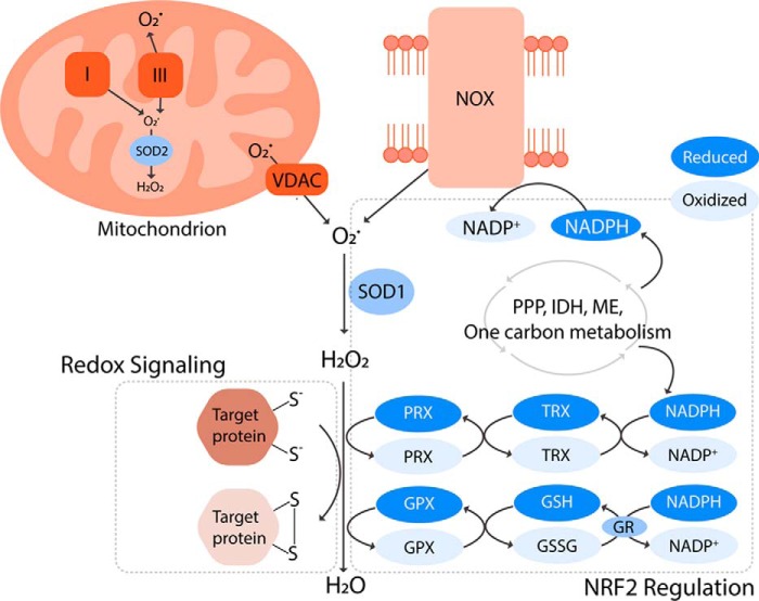
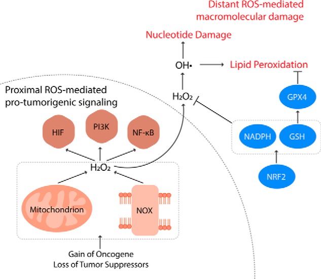
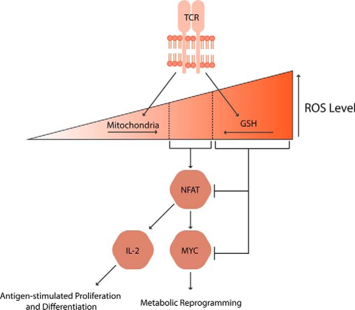
Reactive oxygen species (ROS) mediate redox signaling necessary for numerous cellular functions. Yet, high levels of ROS in cells and tissues can cause damage and cell death. Therefore, regulation of redox homeostasis is essential for ROS-dependent signaling that does not incur cellular damage. Cells achieve this optimal balance by coordinating ROS production and elimination. In this Minireview, we discuss the mechanisms by which proliferating cancer and T cells maintain a carefully controlled redox balance. Greater insight into such redox biology may enable precisely targeted manipulation of ROS for effective medical therapies against cancer or immunological disorders.

Keywords: T-cell; cancer; reactive oxygen species (ROS); redox regulation; redox signaling.


Conflict of interest statement

The authors declare that they have no conflicts of interest with the contents of this article

Figures

Figure 1.
Figure 1.
Production, elimination, and signaling of ROS. Mitochondria and NADPH oxidases (NOXs) are the main sources of superoxide (O2˙̄), which is converted to hydrogen peroxide (H2O2) by superoxide dismutases (SODs). H2O2 can subsequently (a) oxidize thiols within redox-regulated proteins to conduct cellular signaling or (b) be reduced to water by antioxidant systems largely composed of NRF2-regulated enzymes. The peroxiredoxin (PRX)/thioredoxin (TRX) and glutathione peroxidase (GPX)/glutathione (GSH) systems are fueled by NADPH. This key reducing equivalent is generated by a complex network of metabolic pathways and enzymes involving the pentose phosphate pathway (PPP), isocitrate dehydrogenases (IDHs), malic enzymes (MEs), and one-carbon metabolism. Interestingly, NADPH is also a substrate for the ROS-generating NOXs. This suggests that the antioxidant systems and ROS producers are equally important for biological processes, and they work in concert to regulate redox environments that permit the ROS-mediated signaling without incurring oxidative damage.
Figure 2.
Figure 2.
Regulation of redox balance in cancer cells. Compared with nontransformed cells, cancer cells have elevated levels of ROS instigated by acquisition of oncogenes and loss of tumor suppressors. ROS from mitochondria and NOXs oxidize co-localized redox-regulated target proteins to activate pro-tumorigenic signaling pathways, including HIF-1α, PI3K, and NF-κB. Distant from the sites of production, however, ROS nonspecifically react with nucleotides and lipids inducing oxidative damage and even cell death. These distant damaging ROS can be controlled by antioxidant systems such as NRF2, NADPH generation, GSH synthesis/regeneration, and GPX4. Therefore, cancer cells producing elevated levels of ROS concomitantly increase such antioxidant capacities. This shift in redox balance enables cancer cells to hyper-activate the proximal ROS-mediated pro-survival and proliferation signaling without experiencing ROS toxicity.
Figure 3.
Figure 3.
Regulation of redox balance in T cells. Both ROS generation by mitochondria and ROS scavenging by GSH are essential for the T cell activation. The ROS at the defined window activates nuclear factor of activated T cells (NFAT), which in turn induces IL-2 and MYC expressions, leading to T cell metabolic reprogramming, expansion, and differentiation into effector or regulator cells.

References

    1. Holmström K. M., and Finkel T. (2014) Cellular mechanisms and physiological consequences of redox-dependent signaling. Nat. Rev. Mol. Cell Biol. 15, 411–421 10.1038/nrm3801 - DOI - PubMed
    1. Raimundo N., Song L., Shutt T. E., McKay S. E., Cotney J., Guan M. X., Gilliland T. C., Hohuan D., Santos-Sacchi J., and Shadel G. S. (2012) Mitochondrial stress engages E2F1 apoptotic signaling to cause deafness. Cell 148, 716–726 10.1016/j.cell.2011.12.027 - DOI - PMC - PubMed
    1. Chouchani E. T., Pell V. R., Gaude E., Aksentijević D., Sundier S. Y., Robb E. L., Logan A., Nadtochiy S. M., Ord E. N. J., Smith A. C., Eyassu F., Shirley R., Hu C. H., Dare A. J., James A. M., et al. (2014) Ischaemic accumulation of succinate controls reperfusion injury through mitochondrial ROS. Nature 515, 431–435 10.1038/nature13909 - DOI - PMC - PubMed
    1. Brand M. D. (2016) Mitochondrial generation of superoxide and hydrogen peroxide as the source of mitochondrial redox signaling. Free Radic. Biol. Med. 100, 14–31 10.1016/j.freeradbiomed.2016.04.001 - DOI - PubMed
    1. Bedard K., and Krause K. H. (2007) The NOX family of ROS-generating NADPH oxidase: physiology and pathophysiology. Physiol. Rev. 87, 245–313 10.1152/physrev.00044.2005 - DOI - PubMed

Publication types

Substances

LinkOut - more resources