. 2018 Mar 2;293(9):3265-3280.
doi: 10.1074/jbc.M117.816348. Epub 2017 Dec 27.

Exceptionally high-affinity Ras binders that remodel its effector domain

Affiliations

Exceptionally high-affinity Ras binders that remodel its effector domain

John H McGee et al. J Biol Chem. .

Abstract

The Ras proteins are aberrantly activated in a wide range of human cancers, often endowing tumors with aggressive properties and resistance to therapy. Decades of effort to develop direct Ras inhibitors for clinical use have thus far failed, largely because of a lack of adequate small-molecule-binding pockets on the Ras surface. Here, we report the discovery of Ras-binding miniproteins from a naïve library and their evolution to afford versions with midpicomolar affinity to Ras. A series of biochemical experiments indicated that these miniproteins bind to the Ras effector domain as dimers, and high-resolution crystal structures revealed that these miniprotein dimers bind Ras in an unprecedented mode in which the Ras effector domain is remodeled to expose an extended pocket that connects two isolated pockets previously found to engage small-molecule ligands. We also report a Ras point mutant that stabilizes the protein in the open conformation trapped by these miniproteins. These findings provide new tools for studying Ras structure and function and present opportunities for the development of both miniprotein and small-molecule inhibitors that directly target the Ras proteins.

Keywords: Ras protein; X-ray crystallography; cancer; directed evolution; peptides.


Conflict of interest statement

The research reported herein is the subject of a license option agreement between Harvard University and FOG Pharmaceuticals. G. L. V. is a major shareholder in FOG Pharmaceuticals

Figures

Figure 1.
Figure 1.
Discovery of miniproteins that bind the Ras proteins and block the Ras-Raf interaction. A, structure of the aPP scaffold used as the basis of the library with randomized residues shown in orange (Protein Data Bank code 1PPT). B, left panel, FACS plots of representative isolates from each round when incubated with 250 nm KRas(G12V)-GppNHp-Alexa Fluor 647. Right panel, FACS of the 225-3 clone in the presence of 250 nm KRas(G12V)-GppNHp-Alexa Fluor 647 with or without 5 μm unlabeled B-Raf RBD. C, sequences of library members isolated after each round of enrichment. The asterisk (*) indicates Y7C mutation. The initial library screen (to obtain hits) was performed twice and afforded similar hit sequences; the remaining evolution rounds were performed once.
Figure 2.
Figure 2.
Biochemical characterization of Ras-binding miniproteins. A, fluorescence polarization of FITC-labeled 225-3 miniprotein with Ras proteins and Ras family members. All proteins are loaded with GppNHp, and all contain the G12V mutation except for RalA and Rab25 (wildtype). Dissociation constant (Kd) values are mean ± S.E. for experiments performed in triplicate. B, dissociation of mant-labeled GppNHp from KRas(G12V) in the presence of 2 μm ligand, initiated by the addition of 1 mm GppNHp. C, pulldown of endogenous Ras from Capan-1 pancreatic adenocarcinoma lysate by GST-labeled B-Raf RBD after incubation with or without miniprotein. B-Raf RBD and associated proteins were isolated with glutathione beads and separated by SDS-PAGE prior to transfer and Western blotting (WB) with a pan-Ras antibody. The molecular weight ladder lane is spliced to conserve space. D, CD of purified miniproteins at 20–50 μm concentration in 50 mm sodium phosphate, pH 8. E, Kd values of alanine mutagenesis peptides with KRas(G12V)-GppNHp determined as in A. F, SPR of free Ras (or miniproteins) that was flowed over a biotin CAPture sensor chip onto which biotinylated miniprotein (or Ras) (respectively) was immobilized. The experiment was performed in single-cycle format with injections at increasing concentration of free analyte. All experiments were performed at least three times. deg, degrees; RU, resonance units; RFU, relative fluorescence units.
Figure 3.
Figure 3.
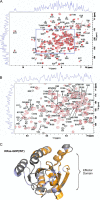
Miniproteins bind the Ras effector domain in solution. A, 1H-15N TROSY correlation data recorded for a sample of 15N-labeled KRas-GDP(WT) in the absence (black) or presence (red) of the 225-1 miniprotein. KRas cross-peaks were identified by comparison with published assignments (26) and used to determine significant chemical shift perturbations on a per-residue basis. B, zoom of the dashed blue boxed region from A. A majority (147 of 161) of the observable residues were identified (excluding the non-observable N terminus and four prolines). Overlap for 15 residues precluded either assignment and/or determination of chemical shift perturbation status. C, mapping of the chemical shift perturbation data on the structure of KRas-GDP(WT) (Protein Data Bank code 4OBE). Residues with significant chemical shift perturbation effects are colored orange, non-perturbed residues are gray, and proline/undetermined residues are light blue. An extensive chemical shift perturbation pattern was observed centered on the switch regions and supporting secondary structure elements. Minor chemical shift perturbation effects were observed in distal regions of the structure; collectively, the data are consistent with a remodeling of the Ras effector–binding region.
Figure 4.
Figure 4.
Miniproteins bind to KRas as a homodimer. A, 1H-15N heteronuclear single quantum correlation nuclear magnetic resonance spectroscopy on 15N-labeled 225-1 miniprotein in the absence or presence of unlabeled KRas(G12V)-GppNHp protein. The 225-1 miniprotein is used in place of 225-3 due to limiting solubility. B, sequence of 225-11 miniproteins and dissociation of mant-GppNHp from KRas(G12V) in the presence of disulfide- or diselenide-bonded 225-11 miniprotein dimers and in the presence or absence of 2 mm DTT as reducing agent. The inset shows reverse-phase HPLC of miniproteins treated with or without 10 mm DTT; the identity of reduced and oxidized species was confirmed by mass spectrometry. U, selenocysteine. HSQC samples were prepared and recorded once; mant dissociation was performed three times. RFU, relative fluorescence units.
Figure 5.
Figure 5.
Miniprotein homodimers bind to the KRas effector domain in an open conformation. A, crystal structure of KRas(G12V)-GppNHp (blue) in complex with the 225-11 miniprotein (green) at 2.3-Å resolution. Switch I and switch II of the effector domain are shown in orange, inter-miniprotein disulfide bond is shown in yellow sticks, nucleotide is represented as sticks, and N and C termini are indicated for miniprotein protomers. B, overlay of KRas(Q61H)-GppNHp alone (white; Protein Data Bank code 3GFT) with the structure from A with miniprotein removed for clarity. C, structures from A and B with surface rendering of KRas. D, crystal structure of KRas(G12V)-GppNHp (blue) in complex with the 225-11 A30R miniprotein mutant (green) at 1.7-Å resolution. GppNHp, disulfide bond, and Arg-30 are represented as sticks, and coordinated waters and Mg2+ ion are represented as spheres.
Figure 6.
Figure 6.
Comparison of Ras surface and nucleotide pocket conformations. A, crystal structure of KRas(G12V)-GppNHp bound to 225-11 or KRas(WT)-apo bound to SOS1. Contact residues on Ras within 4 Å of 225-11 or SOS are highlighted in yellow. B, close-up of nucleotide-binding pocket in KRas(G12V)-GppNHp bound to 225-11 (blue) aligned with KRas(Q61H)-GppNHp (white; PDB 3GFT). Coordinated Mg2+ ions are represented as spheres.
Figure 7.
Figure 7.
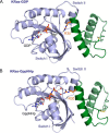
Comparison of 225-11 A30R miniprotein bound to KRas-GppNHp versus KRas-GDP. A, crystal structure of 225-11 A30R miniprotein (green) bound to Ras-GDP (blue) at 2.2-Å resolution. B, crystal structure of 225-11 A30R miniprotein (green) bound to Ras-GppNHp (blue) at 1.7-Å resolution. Switch I was partially disordered in both structures. GDP/GppNHp, disulfide bond, and R30 are represented as sticks, coordinated waters and Mg2+ ion are represented as spheres, and dashed lines indicate distances less than 3 Å.
Figure 8.
Figure 8.
Sequence and structure of a miniprotein heterodimer that binds Ras with improved affinity. A, sequence of heterodimeric miniprotein isolated from YSD screen. Mutated residues relative to 225-11 are shown in orange. B, crystal structure of KRas(G12V)-GppNHp (blue) in complex with the 225-15a/b miniprotein (225-15a protomer in dark green; 225-15b protomer in light green) at 1.7-Å resolution. Mutated residues compared with 225-11 are shown in orange sticks. Switch I is partially disordered in this structure.
Figure 9.
Figure 9.
The D38P mutant stabilizes KRas in an open conformation. A, 31P NMR spectra of GppNHp-bound Ras mutants to determine switch I dynamics. Peaks are assigned based on prior studies of HRas (34). B, dissociation of mant-GppNHp from KRas(G12V) or KRas(G12V,D38P) in the presence of 2 μm ligand, initiated by the addition of 1 mm GTP. C, aligned structures of KRas(G12V)-GppNHp at 2.1-Å resolution and KRas(G12V,D38P)-GppNHp at 1.9-Å resolution (both bound to 225-11) with residue 38 and nearby side chains shown as sticks. D, aligned structures of KRas(G12V)-GDP (Protein Data Bank code 4TQ9) and KRas(G12V,D38P)-GDP at 1.6-Å resolution with Asp-38 and Pro-38 shown as sticks. Switch I is partially disordered for KRas(G12V,D38P)-GDP. NMR spectra were recorded once (∼48-h collection each); mant dissociation assays were performed three times. RFU, relative fluorescence units.
Figure 10.
Figure 10.
Miniprotein binding remodels the Ras effector domain. Surface representation of KRas(G12V)-GppNHp bound to 225-11 and 90° rotation with cutaway to illustrate the extended pocket is shown. Miniprotein is removed for clarity, and miniprotein residues Trp-25 and Tyr-29 are shown as green sticks. This structure was aligned with KRas bound to Acrylamide 16 (17) (Protein Data Bank code 4M22) and KRas bound to Compound 13 (15) (Protein Data Bank code 4EPY) with the corresponding Ras surface removed and Ras-binding ligands colored in magenta or orange, respectively. Portions of Acrylamide 16 overlap with KRas density from the 225-11 complex structure because Acrylamide 16 induces a larger pocket behind switch II.
Figure 11.
Figure 11.
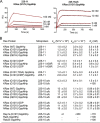
Ras-miniprotein binding data. A, representative SPR sensorgrams. For 225-15a/b, less Ras was immobilized to minimize mass transport limitations, and koff values were only determined from the 3, 10, and 30 nm injections due to an insufficient drop in signal at 1 nm. Labels indicate injected concentration of miniprotein. B, SPR binding parameters determined on a Biacore X100 with a Biotin CAPture kit and biotinylated proteins. Miniproteins were injected at 3, 10, 30, and 100 nm (1, 3, 10, and 30 nm for 225-11 A30R and 225-15a/b). Values are mean ± S.E. of three independent replicates; statistics shown are calculated for kon, koff, and Kd independently.
Figure 12.
Figure 12.
Sequences and masses of miniproteins used in this work and analysis of labeled Ras proteins. A, sequences and calculated/found (electrospray MS, positive mode) masses of recombinantly expressed miniproteins. U, selenocysteine. All dimers are oxidized. B, HPLC traces (A280) of representative miniproteins following purification. C, dissociation of mant-labeled GppNHp from KRas(G12V) proteins in the presence or absence of 5 μm Raf RBD, initiated by the addition of 1 mm GppNHp. D, size-exclusion chromatography of KRas(G12V) proteins (Superdex 200 Increase 10/300 GL column; void volume is at ∼7 ml). RFU, relative fluorescence units.

References

    1. Young A., Lyons J., Miller A. L., Phan V. T., Alarcón I. R., and McCormick F. (2009) Ras signaling and therapies. Adv. Cancer Res. 102, 1–17 10.1016/S0065-230X(09)02001-6 - DOI - PubMed
    1. Karnoub A. E., and Weinberg R. A. (2008) Ras oncogenes: split personalities. Nat. Rev. Mol. Cell Biol. 9, 517–531 10.1038/nrm2438 - DOI - PMC - PubMed
    1. Drosten M., Lechuga C. G., and Barbacid M. (2014) Ras signaling is essential for skin development. Oncogene 33, 2857–2865 10.1038/onc.2013.254 - DOI - PubMed
    1. Prior I. A., Lewis P. D., and Mattos C. (2012) A comprehensive survey of Ras mutations in Cancer. Cancer Res. 72, 2457–2467 10.1158/0008-5472.CAN-11-2612 - DOI - PMC - PubMed
    1. Pylayeva-Gupta Y., Grabocka E., and Bar-Sagi D. (2011) RAS oncogenes: weaving a tumorigenic web. Nat. Rev. Cancer 11, 761–774 10.1038/nrc3106 - DOI - PMC - PubMed

Publication types