. 2018 Feb 1;200(3):928-936.
doi: 10.4049/jimmunol.1700273. Epub 2017 Dec 27.

Inhibition of the Deubiquitinase Usp14 Diminishes Direct MHC Class I Antigen Presentation

Affiliations

Inhibition of the Deubiquitinase Usp14 Diminishes Direct MHC Class I Antigen Presentation

Amy L Palmer et al. J Immunol. .

Abstract

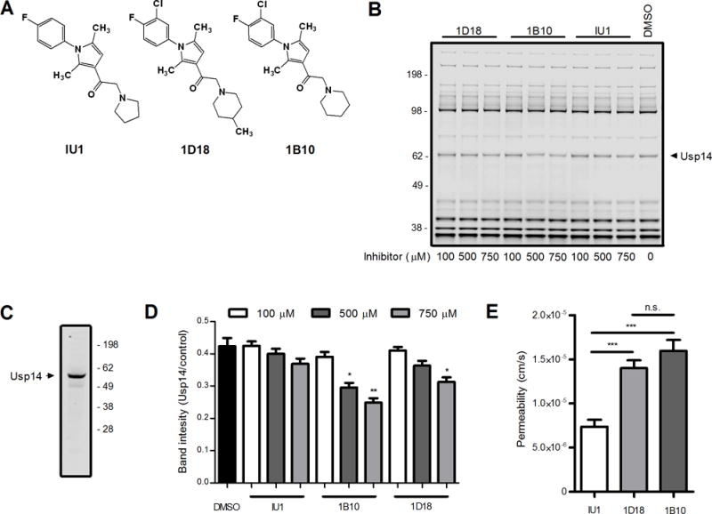
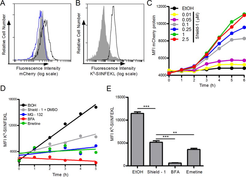
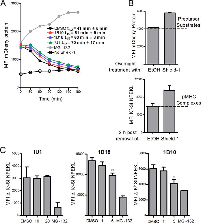
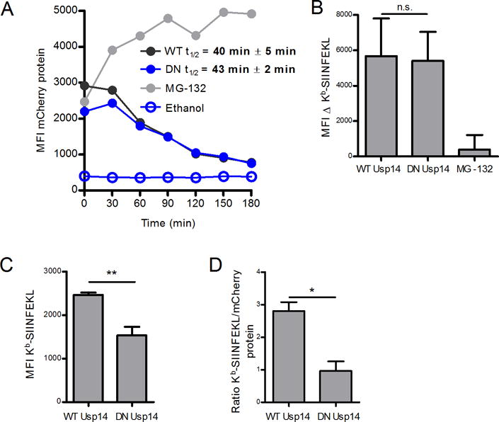
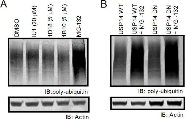
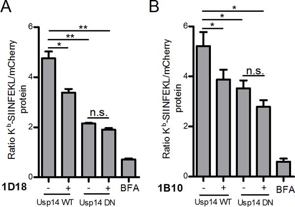
Infected or transformed cells must present peptides derived from endogenous proteins on MHC class I molecules to be recognized and targeted for elimination by Ag-specific cytotoxic T cells. In the first step of peptide generation, proteins are degraded by the proteasome. In this study, we investigated the role of the ubiquitin-specific protease 14 (Usp14), a proteasome-associated deubiquitinase, in direct Ag presentation using a ligand-stabilized model protein expressed as a self-antigen. Chemical inhibition of Usp14 diminished direct presentation of the model antigenic peptide, and the effect was especially pronounced when presentation was restricted to the defective ribosomal product (DRiP) form of the protein. Additionally, presentation specifically from DRiP Ags was diminished by expression of a catalytically inactive form of Usp14. Usp14 inhibition did not appreciably alter protein synthesis and only partially delayed protein degradation as measured by a slight increase in the half-life of the model protein when its degradation was induced. Taken together, these data indicate that functional Usp14 enhances direct Ag presentation, preferentially of DRiP-derived peptides, suggesting that the processing of DRiPs is in some ways different from other forms of Ag.


Figures

Figure 1
Figure 1. Compounds 1D18 and 1B10 are comparable to IU1
Compounds 1D18 and 1B10 were directly assessed against the selective Usp14 inhibitor IU1. A. Schematic depiction of the chemical structures of all three compounds. B. EL4 cell lysates were incubated with each compound of interest at various concentrations indicated for 60 min. Lysates were probed with Rho-Ub-PA and then resolved by SDS-PAGE to assess levels of deubiquitinating activity found throughout the cell. Usp14 is indicated in the figure based upon previous experiments (16) and Western blot of Usp14 (C). D. Analysis of DUB activity was determined by comparing Usp14 band intensity to the band intensity of a control band. E. Permeability (cm/s) was determined by passive diffusion of chemical compounds from one well to another through an artificial membrane after 6 h incubation at room temperature in the dark (* (* p < 0.01, ** p < 0.001, *** p < 0.0001, n.s. = not significant).
Figure 2
Figure 2. Shield-1 prevents the degradation of SCRAP-mCherry in EL4 cells
A. Fluorescent mCherry protein accumulation after 18 h treatment of EL4/SCRAP-mCherry cells with either ethanol (blue trace) or Shield-1 (black trace) compared to parental EL4 cells (shaded). B. Kb-SIINFEKL expressed in SCRAP-mCherry cells first washed in mild acidic buffer (shaded histogram) then treated with ethanol for 5 h (black trace). C. Kinetic accumulation of mCherry protein over 6 h with the addition of various concentrations of Shield-1. D. SCRAP-mCherry cells were treated with vehicle, 1.0 μM Shield-1, MG-132 (10 μM), Brefeldin A (10 μM), or emetine (10 μM) and aliquots of cell suspensions were collected every hour for 5 hours. Cells were stained as in B. A linear regression line of best fit is shown. E. Similar to D above except only one time point (5 hours) was measured in triplicate after cells were acid washed and treated with ethanol, Shield-1, Brefeldin A, or emetine. Analysis was performed by flow cytometry. (** p˂ 0.01, *** p ˂ 0.001)
Figure 3
Figure 3. Chemical inhibition of Usp14 diminished DRiP Kb-SIINFEKL antigenic presentation
EL4/SCRAP-mCherry cells were utilized to monitor the effects of antigen presentation with Usp14 inhibition by IU1, 1D18, and 1B10. A. Cells were acid-washed to remove existing Kb-SIINFEKL complexes and cultured in the presence of Shield-1 or ethanol and various concentrations of IU1, 1D18 and 1B10. After 5 hours of culture, cells were stained in triplicate with 25D-1.16 mAb. Antigen presentation levels from non-DRiP substrates was determined by subtracting the MFI of 25D-1.16 staining of Shield-1 treated cells from the MFI of ethanol-treated cells. (B). Data from (A) is expressed as a percent inhibition of antigen presentation. The range of Kb-SIINFEKL for non-DRiP substrates was determined by subtracting the MFI of 25D-1.16 staining of Shield-1 treated cells from ethanol treated cells. For DRiP substrates (shaded bars) the range was determined by subtracting the MFI of 25D-1.16 staining of BFA treated cells from Shield-1 treated cells. (*p ˂ 0.05, **p ˂ 0.01, ***p ˂ 0.001, n.s. = not significant).
Figure 4
Figure 4. Chemical inhibition of Usp14 with 1D18 and 1B10 reduces retiree protein degradation
EL4/SCARP-mCherry cells were treated with Shield-1 for 18 h and then acid-washed to remove Kb-SIINFEKL complexes and cultured without Shield-1 and in the presence of MG-132 (10 μM) or the Usp14 inhibitors IU1 (20 μM), 1D18 (5 μM), or 1B10 (5 μM) for 3 hours (A) or 2 hours (B and C). A. SCRAP-mCherry protein degradation is shown at the indicated time points and the calculated half-life for the model protein in the presence of each inhibitor is listed. B. Representative graphs demonstrating (top) the amount of precursor substrates from mCherry that contribute to (bottom) the number of Kb-SIINFEKL complexes observed in the retiree antigen presentation model. The difference in MFI of 25D-1.16 staining between cells with retired SCRAP-mCherry (labeled Shield-1) and cells treated with ethanol alone is termed the ΔKb-SIINFEKL C. Usp14 inhibitors IU1, 1D18, and 1B10 at indicated concentrations were added to cells with retired SCRAP-mCherry and the ΔKb-SIINFEKL was determined 2 h post Shield-1 removal. (*p ˂ 0.05, **p ˂ 0.01)
Figure 5
Figure 5. Catalytically inactive dominant negative Usp14 diminishes DRiP Kb-SIINFEKL presentation
A catalytically deficient dominant negative (DN) Usp14 was utilized to measure effects of Usp14 on antigen presentation. A. Shield-1 treatment (1 μM) of EL4 SCRAP-mCherry cells expressing WT or DN Usp14 for 18 h and subsequent Shield-1 removal allowed for the determination of mCherry protein half-life. B. Kb-SIINFEKL differences between Shield-1 and ethanol treatment were measured in either Usp14 WT or DN cells 2 hours following the removal of Shield-1 and compared to MG-132 treated cells. C. Usp14 WT and Usp14 DN cells were first washed with mild acidic buffer then treated with Shield-1 or ethanol and monitored for Kb-SIINFEKL presentation after 5 h. Cells were stained in triplicate to determine Kb-SIINFEKL levels. D. To account for differences in mCherry levels between the cell types, the MFI of Kb-SIINFEKL signal was normalized to mCherry protein accumulation. (*p ˂ 0.05, **p ˂ 0.05, n.s. = not significant).
Figure 6
Figure 6. Poly-ubiquitinated protein levels are not changed upon Usp14 inhibition
Poly-ubiquitinated protein levels of cells treated with either IU1, 1D18, 1B10, or cells containing the dominant negative phenotype for Usp14 were measured by western blot. A. Poly-ubiquitin staining with FK2 monoclonal mouse antibody after 2 h treatment with vehicle, IU1 (20 μM), 1D18 (5 μM), 1B10 (5 μM), or 10 μM MG-132. B. Cell lysates from WT Usp14 and DN Usp14 were untreated or incubated with MG-132 for 2 h then stained for poly-ubiquitin. Actin antibody was used as a loading control for all poly-ubiquitin western blots.
Figure 7
Figure 7. Chemical inhibition of Usp14 in cells containing catalytically inactive Usp14 does not further diminish antigenic peptide presentation
Effects of both Usp14 inhibition, with chemical compounds, and dominant negative form of Usp14 on DRiP antigen presentation was measured. WT Usp14 WT and DN Usp14 cells treated with 5 μM of 1B10 (A) or 1D18 (B) for 5 h and aliquots of cell suspension were analyzed by flow cytometry for the detection of Kb-SIINFEKL complexes and mCherry fluorescence. Kb-SIINFEKL was normalized to mCherry protein accumulation. BFA was added to a portion of cells to act as a positive control for complete reduction of antigen presentation. (p ˂ 0.05 =*, p ˂ 0.01 =**, n.s. = not significant)

Similar articles

Cited by

References

    1. van Endert P. Post-proteasomal and proteasome-independent generation of MHC class I ligands. Cellular and Molecular Life Sciences. 2011;68:1553–1567. - PMC - PubMed
    1. Lee MJ, Lee BH, Hanna J, King RW, Finley D. Trimming of Ubiquitin Chains by Proteasome-associated Deubiquitinating Enzymes. Molecular & Cellular Proteomics. 2011;10 - PMC - PubMed
    1. Kim HM, Yu Y, Cheng Y. Structure characterization of the 26S proteasome. Biochimica et Biophysica Acta (BBA) - Gene Regulatory Mechanisms. 2011;1809:67–79. - PMC - PubMed
    1. Setz C, Friedrich M, Hahn S, Dörrie J, Schaft N, Schuler G, Schubert U. Just One Position-Independent Lysine Residue Can Direct MelanA into Proteasomal Degradation following N-Terminal Fusion of Ubiquitin. PloS one. 2013;8:e55567. - PMC - PubMed
    1. Rodriguez F, Zhang J, Whitton JL. DNA immunization: ubiquitination of a viral protein enhances cytotoxic T-lymphocyte induction and antiviral protection but abrogates antibody induction. Journal of virology. 1997;71:8497–8503. - PMC - PubMed

Publication types

MeSH terms

Substances