

Microvillus Inclusion Disease Variant in an Infant with Intractable Diarrhea
- PMID: 29282386
- PMCID: PMC5731099
- DOI: 10.1159/000479624
Microvillus Inclusion Disease Variant in an Infant with Intractable Diarrhea
Abstract
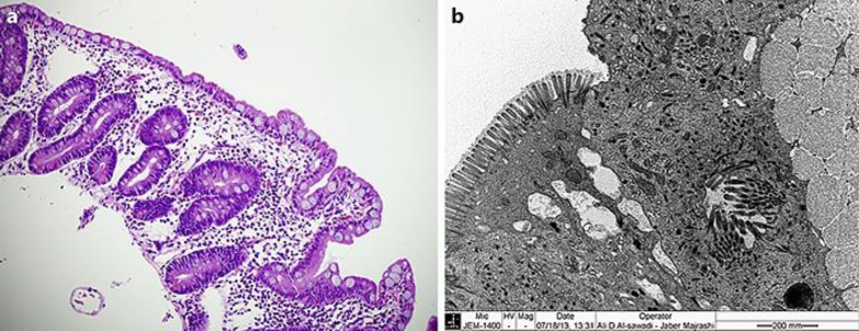
Microvillus inclusion disease (MVID) is a rare autosomal recessive congenital enteropathy characterized by intractable secretory diarrhea. We report a case of MVID variant with a homozygous gene mutation in syntaxin 3 (STX3). The patient is a male Saudi infant who presented shortly after birth with severe vomiting, metabolic acidosis, and mild diarrhea. Electron microscopy study for small intestinal biopsy was consistent with MVID. MYO5B gene mutation was excluded; subsequently, whole exome sequencing (WES) was performed, which revealed homozygous gene mutation in STX3. Using WES in clinical environment can be a useful tool for diagnosing difficult and rare inherited congenital enteropathies.
Keywords: Intractable diarrhea; Microvillus inclusion disease; Syntaxin 3; Whole exome sequencing.
Figures
References
-
- Goulet O, Ruemmele F, Lacaille F, et al. Irreversible intestinal failure. J Pediatr Gastroenterol Nutr. 2004;38:250–269. - PubMed
-
- Sherman PM, Mitchell DJ, Cutz E. Neonatal enteropathies: defining the causes of protracted diarrhea of infancy. J Pediatr Gastroenterol Nutr. 2004;38:16–26. - PubMed
-
- Cutz E, Rhoads JM, Drumm B, et al. Microvillus inclusion disease: an inherited defect of brush-border assembly and differentiation. N Engl J Med. 1989;320:646–651. - PubMed
-
- Phillips AD, Schmitz J. Familial microvillous atrophy: a clinicopathological survey of 23 cases. J Pediatr Gastroenterol Nutr. 1992;14:380–396. - PubMed
Publication types
LinkOut - more resources
Full Text Sources
Other Literature Sources
Molecular Biology Databases
