

Sinonasal Tract Solitary Fibrous Tumor: A Clinicopathologic Study of Six Cases with a Comprehensive Review of the Literature
- PMID: 29282671
- PMCID: PMC6232205
- DOI: 10.1007/s12105-017-0878-y
Sinonasal Tract Solitary Fibrous Tumor: A Clinicopathologic Study of Six Cases with a Comprehensive Review of the Literature
Abstract
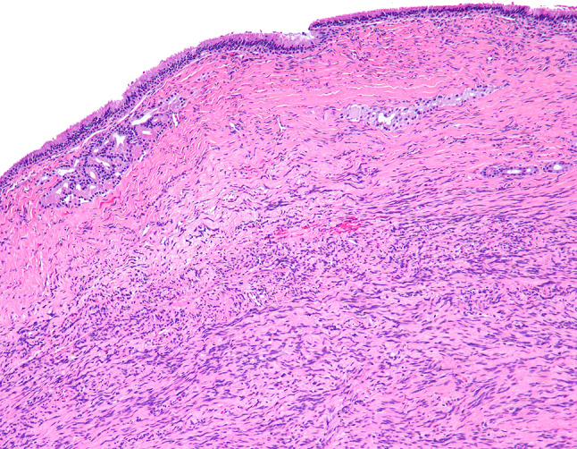
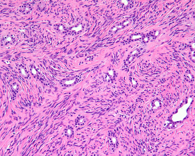
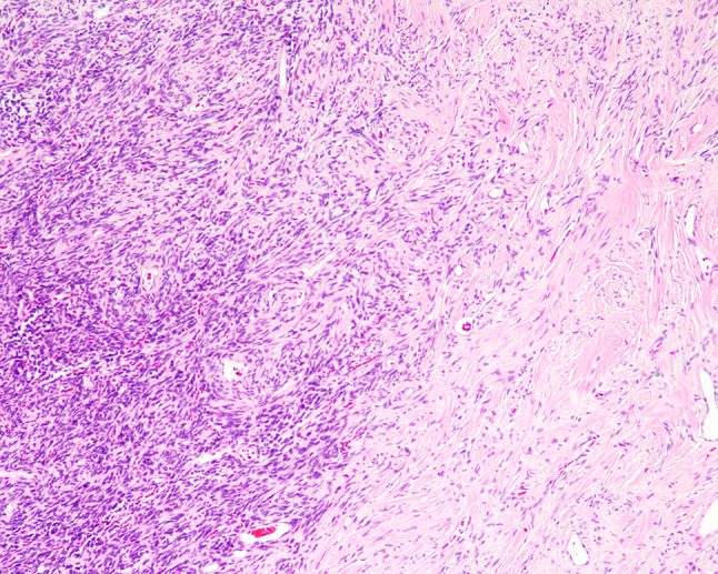
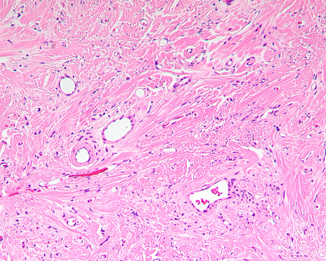
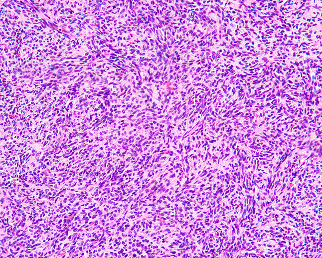
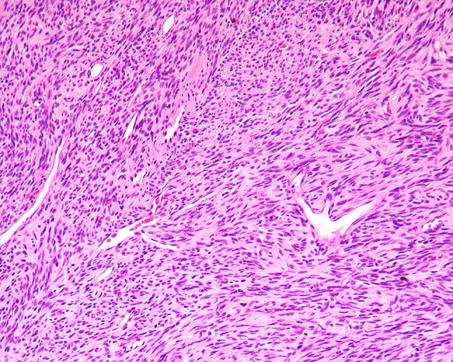
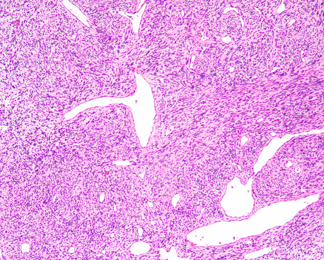
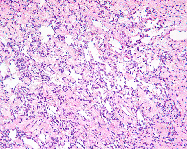
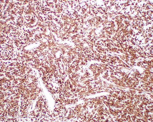
Solitary fibrous tumors (SFTs) are well recognized in the head and neck region, but rarely arise in the sinonasal tract (SNT). Six primary SNT SFTs were identified in the files of Southern California Permanente Medical Group between 2006 and 2017. The patients included five males and one female ranging in age from 33 to 72 years (mean 52 years), most of whom presented clinically with nasal obstruction. Three tumors involved the nasal cavity alone, one involved the paranasal sinuses, and two involved both the nasal cavity and paranasal sinuses. Histologically, the tumors were characterized by a variably cellular proliferation of cytologically bland spindle cells within a collagenous stroma with prominent interspersed branching vessels. Mitotic activity was low (range 0-2 per 10 high power fields) and there was no evidence of pleomorphism or tumor necrosis. Surface ulceration was noted. By immunohistochemistry, the lesional cells were positive for CD34, STAT6 and bcl-2. Clinical follow up information was available for all patients (range 32-102 months; mean 72 months). There were no recurrences or metastases and all were alive with no evidence of disease at last follow-up. SFTs rarely affect the SNT, but should be considered in the differential diagnosis of SNT mesenchymal lesions. Immunohistochemical expression of STAT6 can aid in diagnosis and separation of SFT from other spindle cell lesions occurring at this anatomic site. In combination with cases reported in the literature, primary SNT SFT behave in an indolent manner with conservative treatment.
Keywords: Differential diagnosis; Human; Immunohistochemistry; Nasal cavity; Paranasal sinuses; STAT6 protein; Solitary fibrous tumors.
Conflict of interest statement
All authors declare that they have no conflict of interest as it relates to this research project.
Figures
References
-
- Kao YC, Lin PC, Yen SL, Huang SC, Tsai JW, Li CF, et al. Clinicopathological and genetic heterogeneity of the head and neck solitary fibrous tumours: a comparative histological, immunohistochemical and molecular study of 36 cases. Histopathology. 2016;68(4):492–501. doi: 10.1111/his.12772. - DOI - PubMed
Publication types
MeSH terms
Substances
LinkOut - more resources
Full Text Sources
Other Literature Sources
Medical
Research Materials
Miscellaneous
