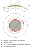
Refractive lenticule extraction small incision lenticule extraction: A new refractive surgery paradigm
- PMID: 29283117
- PMCID: PMC5778540
- DOI: 10.4103/ijo.IJO_761_17
Refractive lenticule extraction small incision lenticule extraction: A new refractive surgery paradigm
Abstract
Small incision lenticule extraction (SMILE), a variant of refractive lenticule extraction technology is becoming increasingly popular, as a flapless and minimally invasive form of laser vision correction (LVC) for the treatment of myopia and myopic astigmatism. This review aims at summarizing the principles, surgical technique, and clinical outcomes in terms of visual and refractive results, safety, efficacy, postoperative dry eye, aberrations, and biomechanics of SMILE and its comparison with other conventional techniques of LVC, such as laser in situ keratomileusis (LASIK) and photorefractive keratectomy (PRK). Recent advancements in the laser frequency and energy delivery patterns, instrumentation, and surgical techniques have shown significant improvement in the visual recovery and outcomes after SMILE, compared to the initial results published by Sekundo and Shah et al. Most of the recently published literature on long-term outcomes of SMILE shows excellent stability of the procedure, especially for higher myopia. In terms of the postoperative dry eye, SMILE shows a clear advantage over LASIK as numerous studies have shown significant differences about the Schirmer's, Tear film break up time, corneal sensitivity, and corneal nerve regeneration to be better following SMILE compared to LASIK. There is some evidence that since the Bowman's membrane (BM) and the anterior lamellae remain intact after SMILE, this may be a potential advantage for corneal biomechanics over LASIK and PRK where the BM is either severed or ablated, respectively, however, the data on biomechanics are inconclusive at present. Overall, this procedure has proved to be promising, delivering equivalent, or better visual and refractive results to LASIK and providing clear advantage in terms of being a flapless, minimally invasive procedure with minimal pain and postoperative discomfort thus offering high patient satisfaction.
Conflict of interest statement
Dr. Sri Ganesh is a consultant for Carl Zeiss Meditec, Germany and receives travel grants from the company.
Figures


References
-
- Sekundo W, Kunert K, Russmann C, Gille A, Bissmann W, Stobrawa G, et al. First efficacy and safety study of femtosecond lenticule extraction for the correction of myopia: Six-month results. J Cataract Refract Surg. 2008;34:1513–20. - PubMed
-
- Sekundo W, Kunert KS, Blum M. Small incision corneal refractive surgery using the small incision lenticule extraction (SMILE) procedure for the correction of myopia and myopic astigmatism: Results of a 6 month prospective study. Br J Ophthalmol. 2011;95:335–9. - PubMed
-
- Krueger RR, Juhasz T, Gualano A, Marchi V. The picosecond laser for nonmechanical laser in situ keratomileusis. J Refract Surg. 1998;14:467–9. - PubMed
-
- Ito M, Quantock AJ, Malhan S, Schanzlin DJ, Krueger RR. Picosecond laser in situ keratomileusis with a 1053-nm nd: YLF laser. J Refract Surg. 1996;12:721–8. - PubMed
-
- Heisterkamp A, Mamom T, Kermani O, Drommer W, Welling H, Ertmer W, et al. Intrastromal refractive surgery with ultrashort laser pulses: In vivo study on the rabbit eye. Graefes Arch Clin Exp Ophthalmol. 2003;241:511–7. - PubMed
Publication types
MeSH terms
LinkOut - more resources
Full Text Sources
Other Literature Sources
Medical

