

doi: 10.4103/ijo.IJO_559_17.
Single-pass four-throw pupilloplasty for angle-closure glaucoma
Affiliations
- PMID: 29283136
- PMCID: PMC5778544
- DOI: 10.4103/ijo.IJO_559_17
Item in Clipboard
Single-pass four-throw pupilloplasty for angle-closure glaucoma
Indian J Ophthalmol.
2018 Jan.
Abstract
Angle-closure glaucoma is characterized by appositional or synechial closure of the anterior chamber angle with glaucomatous field defects that may or may not be associated with a pupillary block. Surgical pupilloplasty with single-pass four-throw technique helps to alleviate the appositional closure along with the breakage of peripheral anterior synechia, thereby increasing the aqueous outflow and decreasing intraocular pressure.
Conflict of interest statement
There are no conflicts of interest.
Figures
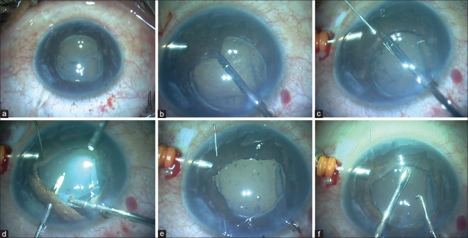
Single-pass 4-throw pupilloplasty technique for angle-closure glaucoma. (a) Cataract extraction done in a case of angle-closure glaucoma. (b) Pupillary stretching. (c) A 10-0 needle passed from proximal iris leaflet. (d) A 26-gauge needle introduced from opposite side through paracentesis incision. (e) The 10-0 needle is docked into 26-gauge needle. (f) Suture loop withdrawn

Single-pass 4-throw pupilloplasty technique for angle-closure glaucoma. (a) Suture end is passed through the loop and 4 throws are taken. (b) Both suture ends are pulled. (c) Knot cut with microscissors. (d) Single-pass four-throw being performed in other quadrant. (e) Both suture ends pulled. (f) Pupillary stretch with 4-point traction

Graphic representation of parameters for all the cases in preoperative and postoperative period. (a) Graph demonstrating area of posterior anterior synechia in the preoperative and postoperative period. (b) Graph demonstrating the degree of peripheral anterior synechia in the pre- and post-operative period. (c) Graph demonstrating the gonioscopy angle grading in pre- and post-operative period

Clinical images with anterior segment optical coherence tomography. (a) A case of angle-closure glaucoma with cataract extraction. (b) Anterior segment optical coherence tomography of angle-closure glaucoma case after cataract extraction. (c) Clinical image of the same case after single-pass four-throw procedure. (d) Anterior segment optical coherence tomography shows open angles with breakage of peripheral anterior synechia and a flat iris plane
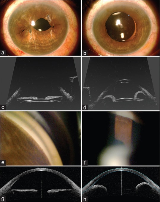
Comparative images of both eyes of a case of angle-closure glaucoma with plateau iris syndrome and cataract extraction (left column = left eye with single-pass four-throw; right column = right eye with no single-pass four-throw). (a) Postoperative image of the left eye with single-pass four-throw and cataract extraction. (b) Postoperative image of the right eye with only cataract extraction done. (c) Ultrasound biomicroscopy denotes open angles with flat iris tissue. (d) Ultrasound biomicroscopy denotes iris bombe with peripheral anterior synechia. (e) Gonioscopy shows open angles. (f) Gonioscopy shows closed angles. (g) Anterior segment optical coherence tomography shows open angles. (h) Anterior segment optical coherence tomography shows angle closure with iris bombe
References
-
- Primary vs. Secondary Angle Closure Glaucoma. American Academy of Ophthalmology. [Last accessed on 2017 Aug 25]. Available from: http://www.eyewiki.aao.org/Primary_vs._Secondary_Angle_Closure_Glaucoma .
-
- Narang P, Agarwal A. Single-pass four-throw technique for pupilloplasty. Eur J Ophthalmol. 2017;27:506–8. - PubMed
-
- Luo M, Liang N. A report of pupilloplasty for secondary glaucoma after vitrectomy associated with ocular trauma. Eye Sci. 2012;27:109–12. - PubMed
-
- Shah M, Law G, Ahmed II. Glaucoma and cataract surgery: Two roads merging into one. Curr Opin Ophthalmol. 2016;27:51–7. - PubMed
-
- He M, Friedman DS, Ge J, Huang W, Jin C, Cai X, et al. Laser peripheral iridotomy in eyes with narrow drainage angles: Ultrasound biomicroscopy outcomes. The Liwan eye study. Ophthalmology. 2007;114:1513–9. - PubMed
Publication types
MeSH terms
LinkOut - more resources
Full Text Sources
Other Literature Sources
Medical
