. 2018 Jan;83(1):153-165.
doi: 10.1002/ana.25130.

Homozygous boricua TBCK mutation causes neurodegeneration and aberrant autophagy

Affiliations

Homozygous boricua TBCK mutation causes neurodegeneration and aberrant autophagy

Xilma R Ortiz-González et al. Ann Neurol. 2018 Jan.

Abstract

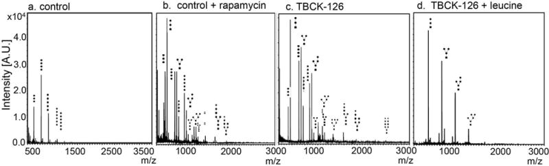
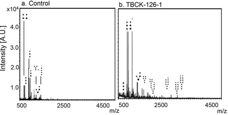
Objective: Autosomal-recessive mutations in TBCK cause intellectual disability of variable severity. Although the physiological function of TBCK remains unclear, loss-of-function mutations are associated with inhibition of mechanistic target of rapamycin complex 1 (mTORC1) signaling. Given that mTORC1 signaling is known to regulate autophagy, we hypothesized that TBCK-encephalopathy patients with a neurodegenerative course have defects in autophagic-lysosomal dysfunction.

Methods: Children (n = 8) of Puerto Rican (Boricua) descent affected with homozygous TBCK p.R126X mutations underwent extensive neurological phenotyping and neurophysiological studies. We quantified autophagosome content in TBCK-/- patient-derived fibroblasts by immunostaining and assayed autophagic markers by western assay. Free sialylated oligosaccharide profiles were assayed in patient's urine and fibroblasts.

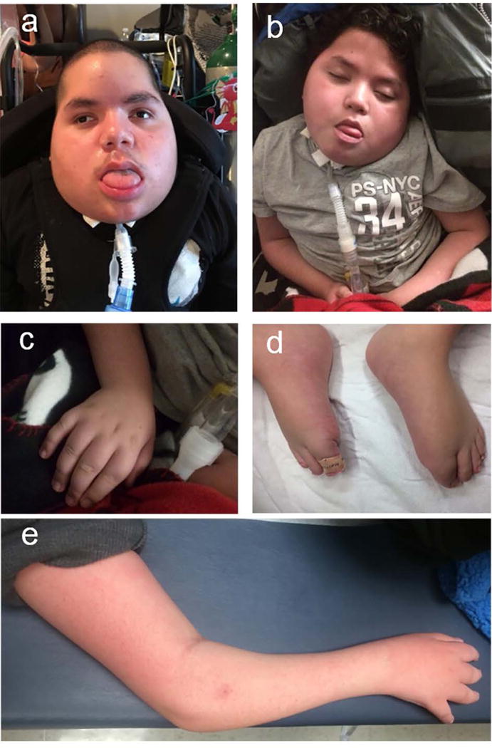
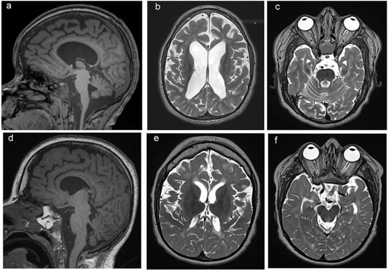
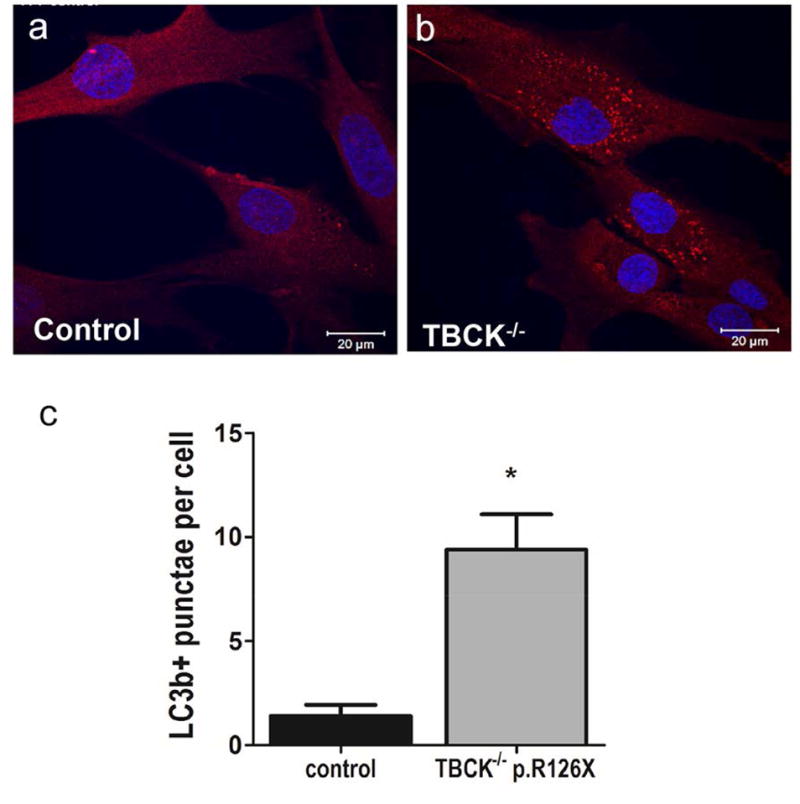
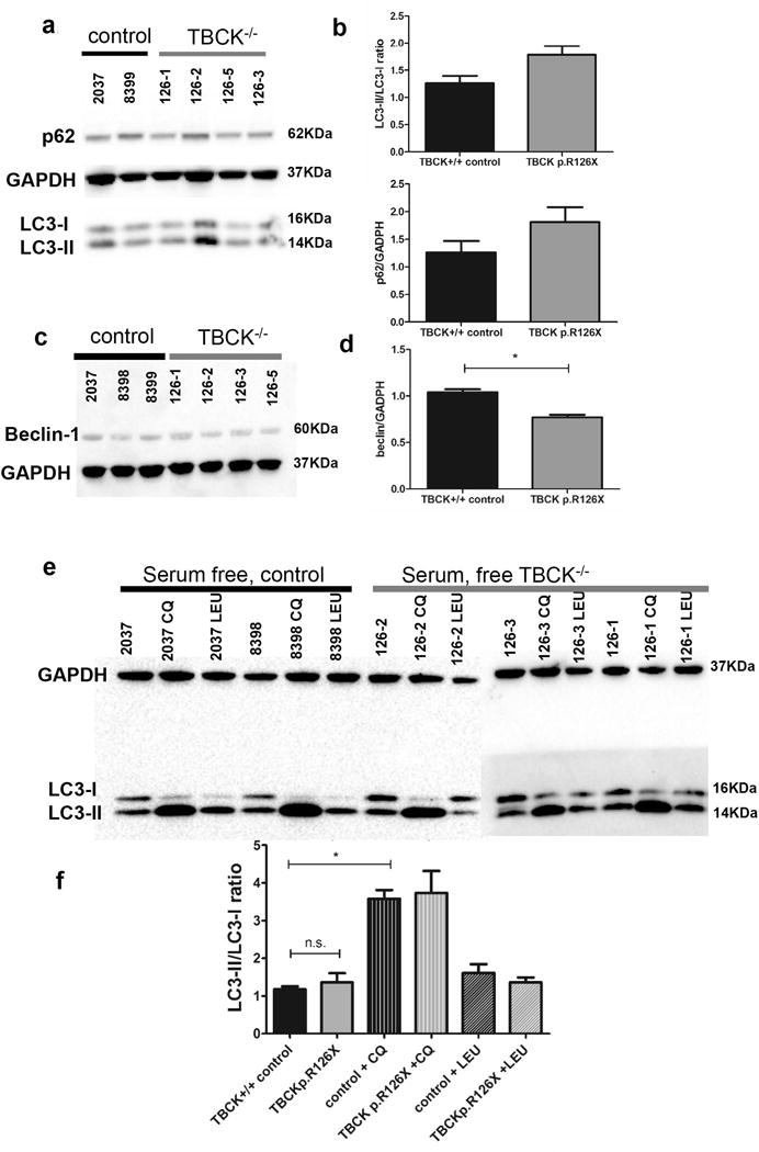
Results: The neurological phenotype of children with TBCK p.R126X mutations, which we call TBCK-encephaloneuronopathy (TBCKE), include congenital hypotonia, progressive motor neuronopathy, leukoencephalopathy, and epilepsy. Systemic features include coarse facies, dyslipidemia, and osteoporosis. TBCK-/- fibroblasts in vitro exhibit increased numbers of LC3+ autophagosomes and increased autophagic flux by immunoblots. Free oligosaccharide profiles in fibroblasts and urine of TBCKE patients differ from control fibroblasts and are ameliorated by treatment with the mTORC1 activator leucine.

Interpretation: TBCKE is a clinically distinguishable syndrome with progressive central and peripheral nervous system dysfunction, consistently observed in patients with the p.R126X mutation. We provide evidence that inappropriate autophagy in the absence of cellular stressors may play a role in this disorder, and that mTORC1 activation may ameliorate the autophagic-lysosomal system dysfunction. Free oligosaccharide profiles could serve as a novel biomarker for this disorder as well as a tool to evaluate potential therapeutic interventions. Ann Neurol 2018;83:153-165.


Conflict of interest statement

Potentials Conflicts of Interest:

Nothing to disclose.

Figures

Figure 1
Figure 1. Clinical Features of TBCK-encephaloneuronopathy
Coarse facial features in patients 126-3 (panel a, age 13 years old) and 126-4 (panel b, age 12 years old); tapered fingers (c) and distal muscle wasting (e). Upper and lower extremity limbs were grossly brachymelic and hands and feet demonstrated acromelia (c, d).
Figure 2
Figure 2
Neuroimaging features of patients with TBCK p.R126X demonstrate thinning of corpus callosum (a, d) and white matter with abnormal T2 signal hyperintensity, most evident in periventricular white matter (b, e) and prominent fourth ventricle due to cerebellar atrophy (c, f). Top panel (a-c): Neuroimaging corresponding to patient 126-3 at age 11years old, bottom panel (d-f), patient 126-2 at age 14 years old.
Figure 3
Figure 3. TBCK−/− fibroblasts show significant increase in LC3+ autophagosomes
LC3b (red) immunostaining of TBCK-encephalopathy patients (b) compared to controls (a). Nuclei stained with DAPI (blue). Panel C shows automated punctae quantification using Zeiss Zen blue software (n=51 wild type and n=106 TBCK cells from 2 independent experiments); bars show SEM, Mann Whitney test p value .0044. TBCK p.R126X data pooled from 3 different patient lines (TBCK 126-1, 126-3, 126-5).
Figure 4
Figure 4. Western Assays from TBCK−/− fibroblasts show increased autophagic flux at baseline but no difference in response to stress or autophagy modulators
Immunoblots for TBCK p.R126X fibroblasts at baseline (complete media) conditions assaying for autophagy markers LC3-I, LC3-II and p62 (panel a) were quantified and normalized to GADPH (panel b). LC3-II/LC3-I ratios are reported as indicative of autophagic flux. Similarly, Beclin-1 levels were assayed (panel c) and quantified relative to GADPH (panel d) at baseline conditions. Quantification includes 3 independent experiments, each including at least 2 wild type and 3 TBCK−/− lines, each blot was normalized to GADPH and statistical significance was defined as two-tailed p value of <.05 using unpaired t-test. To determine if loss-of-function of TBCK affects autophagic response under stress, cells were assayed after 24 hours without serum. Cells were also treated with known autophagy modulators chloroquine (impairs lysosomal function, blocking progression of autophagy) and leucine (enhances mTORC1 signaling). Panel e shows results of immunoblots. As seen in panel f, there is no significant change in the response of controls versus TBCK p.R126X cells under stress or autophagy modulating conditions.
Figure 5
Figure 5. Fibroblasts from TBCK p. R126X patients show aberrant oligosaccharide profiles
MALDI-TOF mass spectrometry profiles of control primary fibroblasts (a). Oligosaccharide species with higher molecular weight (right of profile) accumulate due to impaired autophagic-lysosomal degradation in control cells treated with mTORC1 inhibitor rapamycin (b) and in TBCK−/− R126X cells (c). Degradation defects in TBCK−/− p.R126X cells can be ameliorated by leucine (d), which activates mTORC1 signaling
Figure 6
Figure 6. A representative urine free oligosaccharide profile from a control and a TBCKE patient
Moderate increase of sialylated complexed oligosaccharides were detected in urine from the TBCKE patient (panel b, 126-1) relative to healthy control (panel a), in a pattern consistent with a deficiency in lysosomal associated degradation of glycoproteins.

References

    1. Chong JX, Caputo V, Phelps IG, et al. Recessive Inactivating Mutations in TBCK, Encoding a Rab GTPase-Activating Protein, Cause Severe Infantile Syndromic Encephalopathy. Am J Hum Genet. 2016 Apr;98(4):772–81. - PMC - PubMed
    1. Bhoj EJ, Li D, Harr M, et al. Mutations in TBCK, Encoding TBC1-Domain-Containing Kinase, Lead to a Recognizable Syndrome of Intellectual Disability and Hypotonia. Am J Hum Genet. 2016 Apr;98(4):782–8. - PMC - PubMed
    1. Guerreiro RJ, Brown R, Dian D, de Goede C, Bras J, Mole SE. Mutation of TBCK causes a rare recessive developmental disorder. Neurol Genet. 2016 Jun;2(3):e76. - PMC - PubMed
    1. Mandel H, Khayat M, Chervinsky E, Elpeleg O, Shalev S. TBCK-related intellectual disability syndrome: Case study of two patients. Am J Med Genet A. 2017 Feb;173(2):491–4. - PubMed
    1. Hartley T, Wagner JD, Warman-Chardon J, et al. Whole-exome sequencing is a valuable diagnostic tool for inherited peripheral neuropathies: outcomes from a cohort of 50 families. Clin Genet. 2017 Jul 14; - PubMed

Publication types

MeSH terms