. 2017 Dec 28;12(12):e0189341.
doi: 10.1371/journal.pone.0189341. eCollection 2017.

Protection by extra virgin olive oil against oxidative stress in vitro and in vivo. Chemical and biological studies on the health benefits due to a major component of the Mediterranean diet

Affiliations

Protection by extra virgin olive oil against oxidative stress in vitro and in vivo. Chemical and biological studies on the health benefits due to a major component of the Mediterranean diet

Miriam Rossi et al. PLoS One. .

Abstract

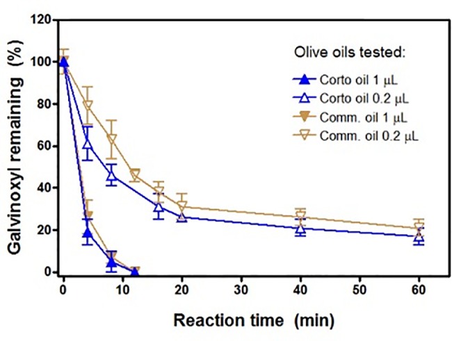
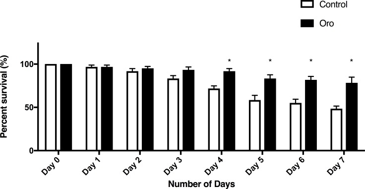
We report the results of in vivo studies in Caenorhabditis elegans nematodes in which addition of extra virgin olive oil (EVOO) to their diet significantly increased their life span with respect to the control group. Furthermore, when nematodes were exposed to the pesticide paraquat, they started to die after two days, but after the addition of EVOO to their diet, both survival percentage and lifespans of paraquat-exposed nematodes increased. Since paraquat is associated with superoxide radical production, a test for scavenging this radical was performed using cyclovoltammetry and the EVOO efficiently scavenged the superoxide. Thus, a linear correlation (y = -0.0838x +19.73, regression factor = 0.99348) was observed for superoxide presence (y) in the voltaic cell as a function of aliquot (x) additions of EVOO, 10 μL each. The originally generated supoeroxide was approximately halved after 10 aliquots (100 μL total). The superoxide scavenging ability was analyzed, theoretically, using Density Functional Theory for tyrosol and hydroxytyrosol, two components of EVOO and was also confirmed experimentally for the galvinoxyl radical, using Electron Paramagnetic Resonance (EPR) spectroscopy. The galvinoxyl signal disappeared after adding 1 μL of EVOO to the EPR cell in 10 minutes. In addition, EVOO significantly decreased the proliferation of human leukemic THP-1 cells, while it kept the proliferation at about normal levels in rat L6 myoblasts, a non-tumoral skeletal muscle cell line. The protection due to EVOO was also assessed in L6 cells and THP-1 exposed to the radical generator cumene hydroperoxide, in which cell viability was reduced. Also in this case the oxidative stress was ameliorated by EVOO, in line with results obtained with tetrazolium dye reduction assays, cell cycle analysis and reactive oxygen species measurements. We ascribe these beneficial effects to EVOO antioxidant properties and our results are in agreement with a clear health benefit of EVOO use in the Mediterranean diet.


Conflict of interest statement

Competing Interests: The authors have declared that no competing interests exist.

Figures

Fig 1
Fig 1. 2-D formal structure of hydroxytyrosol (R: OH) and tyrosol (R: H), left.
Geometry optimized hydroxytyrosol (center) and tyrosol (right) using Dmol3 DFT program; OH groups highlighted in thicker stick mode.
Fig 2
Fig 2. The initial state for reaction (1) between hydroxytyrosol (left) or tyrosol (right) with superoxide radical O2−• obtained through geometry minimization.
Initial separation between O(superoxide) and H was set at 2.60 Å, the sum of van der Waals radii.
Fig 3
Fig 3. The final state of reaction (1) starting from van der Waals separated hydroxytirosol (left) or tyrosol (right) and O2H (2.60 Å), obtained through geometry minimization.
Fig 4
Fig 4. RRDE study of dioxygen in DMSO using gold disk, spinning at 500 rpm, and gold ring electrodes.
The potentials were referenced to a Ag/AgCl and the disk was scanned at 25 mV/sec. The coincidence between forward and reverse potential sweep at both electrodes indicates reversibility of this process.
Fig 5
Fig 5. Ring current of the rotating ring disc electrode system showing the decrease in the oxidation current for the oxidation of superoxide ion radical with the addition of olive oil aliquots.
The experiment consists of sweeping the disk potential from 0.10 V to -1.50 V and then back to 0.10 V while holding the ring potential at -0.25 volts, sufficiently positive to oxidize superoxide back to O2.
Fig 6
Fig 6. Correlation for superoxide scavenging at the ring electrode due to aliquots of olive oil, as indicated in Fig 5.
Collection efficiency is calculated detracting the current at the ring electrode for a given aliquot, at -1.5 V in Fig 5, and the current at time 0, when there was no superoxide yet generated.
Fig 7
Fig 7. Time-course of galvinoxyl disappearance.
Experiments were conducted with 1 μl oil as shown in Fig 8 (1 μl oil to 10 μM galvinoxyl in 100 μl DMSO) and with 0.2 μl oil (1μl oil to 500 μl DMSO) with 10 μM galvinoxyl. Results are the mean ± SD of n = 3 different experiments. Comm. = commercial oil.
Fig 8
Fig 8. Olive oil enhances survival.
L4 nematodes were placed on NGM plates made without or with olive oil, ten nematodes per plate. Survival was scored each day, with surviving worms moved to a fresh treatment plate each day. Two-way ANOVA revealed a significant effect of Oro oil, with greater survival in the presence of the oil than in untreated plates (p<0.001). Bonferroni test for multiple comparisons showed the significant enhancement at days 4–7.
Fig 9
Fig 9. Olive oil enhances survival after paraquat exposure.
L4/adult nematodes were placed on NGM plates with 4mM paraquat with or without olive oil in the nematode growth medium. Nematodes were moved to new treated or untreated plates daily for seven days and the percent of 10 worms on each of six replicate plates for each condition was calculated. Data are the average percent survival across the replicate conditions. Two-way ANOVA for treatment and time exposed revealed a significant effect of treatment and of time, as well as a significant interaction effect (p< 0.001). * significantly different from control survival.
Fig 10
Fig 10. Proliferation curves in the presence of Corto and Oro oils for L-6 myoblasts (upper panel) and THP-1 (lower panel).
Cells were seeded in 60 × 15 mm Petri dishes in 4 ml of the respective medium at appropriate density to reach confluence (or maximum density for the THP-1 cells growing in suspension) after 96 h. Cells were stimulated with each oil 24 h from seeding and then counted every 24 h. Results are the mean ± SD of 3 different experiments carried out in duplicate. *p< 0.001, at least, vs its own control for both 48 and 72 h.
Fig 11
Fig 11. MTT assay carried out with the Corto and Oro olive oils in L6 myoblasts.
Cytotoxicity of cumene hydroperoxide 27.5μM in the presence of Corto and Oro oils at the two concentration used throughout the experiments. *p<0.05, at least, vs all.
Fig 12
Fig 12. Time course of the capability of Oro oil to protect toward oxidative stress given by Cumene hydroperoxide in L6 myoblasts and THP-1 monocytes.
For the L6 myoblasts (upper panel) the concentration of Cumene hydroperoxide was 27.5 μM [18] and 200 μM for the THP-1 monocytes (lower panel). Before addition of cumene hydroperoxide, cells were pre-incubated with oils at 37°C for 10 min, as reported in the Materials and Methods Section. The results are mean of at least two different experiments carried out in duplicate on 12 wells plates. Upper panel: L6 myoblasts with Oro, 10 min * p<0.05 at least, vs Ctrl, Oro ld, Oro hd, Cum + Oro hd; 1 h, * p<0.05 at least vs Ctrl, Oro ld, Oro hd, cum + Oro ld, 3h * p<0.001 at least.
Fig 13
Fig 13. Flow cytometric analysis of DNA after incubation of L-6 myoblasts with Oro oil for 24 h with low dose and high dose as reported under materials and methods.
The histogram shows a DNA content distribution of propidium iodide fluorescence of one representative experiment. In the right panel is shown the sub-diploid peak due to DNA apoptosis fragmentation of nuclei.
Fig 14
Fig 14. The histogram shows a representative experiment as the percentage of cells cycle phases G1, S and G2/M represent the different phases of the cell cycle determined through the application of electronic markers.
In the lower panels is shown the percentage of apoptotic cells found in Sub G1 at different times and doses of Oro oil.

References

    1. Besnard G, Khadari B, Navascues M, Fernandez-Mazuecos M, El Bakkali A, Arrigo N, et al. (2013) The complex history of the olive tree: from Late Quaternary diversification of Mediterranean lineages to primary domestication in the northern Levant. Proc Biol Sci 280: 20122833 doi: 10.1098/rspb.2012.2833 - DOI - PMC - PubMed
    1. Efe R, Soykan A, Cürebal I, Sönmez S.“Olive and Olive Oil Culture in the Mediterranean Basin”, in Efe R., Öztürk M., Ghazanfar S. (Eds.) Environment and Ecology in the Mediterranean Region, I. Chapter 5, pp.54–64. (2012) Cambridge Scholars Publishing.
    1. Monasterio RP, Fernandez M, Silva MF. (2013) Olive oil by capillary electrophoresis: Characterization and genuineness. J Agric Food Chem 61: 4477–4496. doi: 10.1021/jf400864q - DOI - PubMed
    1. Franco MN, Galeano-Diaz T, Lopez O, Fernandez-Bolanos JG, Sanchez J, De Miguel C, et al. (2014) Phenolic compounds and antioxidant capacity of virgin olive oil. Food Chemistry 163: 289–298. doi: 10.1016/j.foodchem.2014.04.091 - DOI - PubMed
    1. Frankel EN. (2011) Nutritional and biological properties of extra virgin olive oil. J. Agric Food Chem 59: 785–792. doi: 10.1021/jf103813t - DOI - PubMed

Publication types