. 2018 Feb 5;150(2):293-306.
doi: 10.1085/jgp.201711879. Epub 2017 Dec 28.

Junctional trafficking and restoration of retrograde signaling by the cytoplasmic RyR1 domain

Affiliations

Junctional trafficking and restoration of retrograde signaling by the cytoplasmic RyR1 domain

Alexander Polster et al. J Gen Physiol. .

Abstract

The type 1 ryanodine receptor (RyR1) in skeletal muscle is a homotetrameric protein that releases Ca2+ from the sarcoplasmic reticulum (SR) in response to an "orthograde" signal from the dihydropyridine receptor (DHPR) in the plasma membrane (PM). Additionally, a "retrograde" signal from RyR1 increases the amplitude of the Ca2+ current produced by CaV1.1, the principle subunit of the DHPR. This bidirectional signaling is thought to depend on physical links, of unknown identity, between the DHPR and RyR1. Here, we investigate whether the isolated cytoplasmic domain of RyR1 can interact structurally or functionally with CaV1.1 by producing an N-terminal construct (RyR11:4300) that lacks the C-terminal membrane domain. In CaV1.1-null (dysgenic) myotubes, RyR11:4300 is diffusely distributed, but in RyR1-null (dyspedic) myotubes it localizes in puncta at SR-PM junctions containing endogenous CaV1.1. Fluorescence recovery after photobleaching indicates that diffuse RyR11:4300 is mobile, whereas resistance to being washed out with a large-bore micropipette indicates that the punctate RyR11:4300 stably associates with PM-SR junctions. Strikingly, expression of RyR11:4300 in dyspedic myotubes causes an increased amplitude, and slowed activation, of Ca2+ current through CaV1.1, which is almost identical to the effects of full-length RyR1. Fast protein liquid chromatography indicates that ∼25% of RyR11:4300 in diluted cytosolic lysate of transfected tsA201 cells is present in complexes larger in size than the monomer, and intermolecular fluorescence resonance energy transfer implies that RyR11:4300 is significantly oligomerized within intact tsA201 cells and dyspedic myotubes. A large fraction of these oligomers may be homotetramers because freeze-fracture electron micrographs reveal that the frequency of particles arranged like DHPR tetrads is substantially increased by transfecting RyR-null myotubes with RyR11:4300 In summary, the RyR1 cytoplasmic domain, separated from its SR membrane anchor, retains a tendency toward oligomerization/tetramerization, binds to SR-PM junctions in myotubes only if CaV1.1 is also present and is fully functional in retrograde signaling to CaV1.1.


Figures

Figure 1.
Figure 1.
Confocal scans near the surface of myotubes indicate that RyR11:4300 is able to localize at junctions between the sarcoplasmic reticulum and PM only when CaV1.1 is present. (A) After expression in either dysgenic (CaV1.1-null) myotubes (top) or dyspedic (RyR1-null) myotubes (bottom), EYFP-RyR1 displayed a punctate distribution consistent with its targeting to SR–PM junctions. (B) In contrast, EYFP-RyR11:4300 was diffusely distributed in dysgenic myotubes (top, n indicates the nucleus) but was arrayed in discrete foci in dyspedic myotubes (bottom). (C) The lack of fluorescent puncta observed for EYFP-RyR11:4300 in dysgenic myotubes (shown in B, top) was not because the endogenous RyR1 occluded junctional binding sites because diffuse fluorescence was also observed when EYFP-RyR11:4300 was expressed in Cav1.1/RyR1 double-null myotubes. Bars, 5 µm.
Figure 2.
Figure 2.
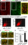
RyR11:4300 colocalizes with CaV1.1. (A) Coexpression of ECFP-CaV1.1 (red) and EYFP-RyR11:4300 (green) in dyspedic myotubes revealed extensive colocalization of the two proteins (yellow-bordered rectangle on the right illustrates the overlay of regions indicated by white rectangles), indicating that the subcellular localization of RyR11:4300 corresponds to junctions containing CaV1.1. Bar, 5 µm. (B) Representative pixel-by-pixel plots of cyan versus yellow fluorescence intensities for images of dyspedic myotubes expressing ECFP-CaV1.1 plus either EYFP-RyR11:4300 (left) or EYFP-RyR1 (right). Individual pixels are represented by a dot with ordinate and abscissa determined by the ECFP and EYFP fluorescence intensity, respectively, of that pixel. (C) Subregions of images obtained from dyspedic myotubes coexpressing ECFP-CaV1.1 and either EYFP-RyR11:4300 (left) or EYFP-RyR1 (right) before (original) and after the ECFP scan (red) was shifted rightward by four pixels (shifted) relative to the EYFP scan (green). Bars, 1 µm. (D) PC values (mean ± SEM) calculated for original and shifted images of dyspedic myotubes expressing ECFP-CaV1.1 and either EYFP-RyR11:4300 or EYFP-RyR1. Seven myotubes were analyzed for both construct combinations, with the PC for an individual myotube determined as the (mean) value for one to three subregions.
Figure 3.
Figure 3.
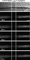
EYFP-RyR11:4300 is mobile within the cytoplasm of dysgenic myotubes. EYFP-RyR11:4300-expressing dysgenic myotubes were selected on the basis of being very long (compared with the width) and of having relatively uniform geometry along their length (top, transmitted light image). The fluorescence within a longitudinal segment near the center (vertical white lines) was bleached by repeated scanning for 5 s at 515 nm with maximum power, followed by imaging at repeated intervals (15, 21, 33, 47, and 111 s postbleach, in this example). Bars, 10 µm.
Figure 4.
Figure 4.
EYFP-RyR11:4300 is stably bound at CaV1.1-containing junctions in dyspedic myotubes. The transmitted light image (top left) shows a suction pipette pressed against the surface of a dyspedic myotube that had been transfected with EGFP-RyR11:4300. The fluorescence images acquired at the indicated times after the beginning of membrane rupture reveal the loss of diffuse green fluorescence and persistence of green fluorescence localized in foci. Bar, 10 µm.
Figure 5.
Figure 5.
Expression of EYFP-RyR11:4300 in dyspedic myotubes produces retrograde enhancement of Ca2+ current equivalent to that of full-length RyR1. (A) Representative L-type Ca2+ currents (depolarizations to −20, 0, +20, and +40 mV) in a nontransfected dyspedic myotube and in dyspedic myotubes expressing either EYFP-RyR1 or EYFP-RyR11:4300. (B) Mean ± SEM peak I-V relationships for dyspedics (n = 15) and dyspedic myotubes expressing EYFP-RyR1 (n = 11) or EYFP-RyR11:4300 (n = 11). Smooth curves represent plots of Eq. 2.
Figure 6.
Figure 6.
EYFP-RyR11:4300 causes a slowing of L-type current activation similar to that caused by EYFP-RyR1. (A–C) Maximal Ca2+ currents in dyspedic myotubes that were nontransfected (white bars, n = 6) or transfected with EYFP-RyR1 (gray bars, n = 10) or EYFP-RyR11:4300 (black bars, n = 10) were fitted as the sum of two exponential functions (Eq. 3), having amplitudes (mean ± SEM) Aslow and Afast (A) and time constants τslow (B) and τfast (C).
Figure 7.
Figure 7.
FPLC indicates that RyR11:4300 in diluted cytosolic lysate of tsA201 cells is partially oligomerized but predominantly monomeric. Lysate from tsA201 cells transfected with EYFP-RyR11:4300 were subjected to FPLC on a Superose 6 10/300 GL column, with the content of EYFP-RyR11:4300 in 0.25-ml eluate fractions determined in triplicate by immunoblotting with monoclonal antibody 34C. The normalized immunostaining intensity is compared with the normalized 280-nm absorbance for the indicated standards. The position of the major immunostaining peak corresponds to that for the predicted mass of monomeric EYFP-RyR11:4300 (512.3 kD), but higher molecular mass oligomers were also present. Error bars represent mean ± SEM.
Figure 8.
Figure 8.
Intermolecular FRET indicates that RyR11:4300 oligomerizes at SR–PM junctions. (A and B) To measure FRET, four confocal scans (S1, S2, S3, and S4) were taken of RyR1/RyR3 double-null myotubes expressing 1:1 mixtures of cDNAs either encoding ECFP-RyR1 and EYFP-RyR1 (A) or encoding ECFP-RyR11:4300 and EYFP-RyR11:4300 (B), in which EYFP was photobleached between S2 and S3. Bars, 5 µm. To measure the cyan fluorescence intensities only within the fluorescent foci, a digital mask was produced by application of an adjustable threshold to the S1 image of EYFP such that all pixel values less than or equal to the threshold were set to zero (shown as black), and all values greater than the threshold (i.e., the majority of the yellow puncta) were set to one (shown as white). (C and D) Pixel intensities for cyan fluorescence were then measured only for mask pixels having the value one, yielding the mean cyan intensity (C1, C2, C3, and C4) for each of the four scans (S1 to S4), as shown in C and D for the cells illustrated in A and B, respectively (values normalized to C1). To account for the unavoidable bleaching of ECFP that occurred during each scan, extrapolated values of cyan fluorescence were calculated as CF = C2 × (C2/C1) and CR = C3 × (C3/C4), which provided two estimates of FRET efficiency for that cell: [C3 − CF]/C3 and [CR − C2]/CR. The mean of these two estimates was taken as the FRET efficiency for the cell, which was 8.5% for the illustrated cell expressing ECFP-RyR1 and EYFP-RyR1 and 3.9% for the cell expressing ECFP-RyR11:4300 and EYFP-RyR11:4300.
Figure 9.
Figure 9.
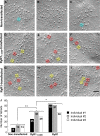
RyR11:4300 causes arrangement of DHPRs into tetrads. (A–I) Representative freeze-fracture replicas are shown of junctional regions of R1R3 myotubes (null for both RyR1 and RyR3) that were either nontransfected (A–C) or transfected with EYFP-RyR11:4300 (D–F) or with EYFP-RyR1 (G–I). Several clusters in the transfected myotubes (D–I) that appear to be partial (three-particle) or complete (four-particle) tetrads are indicated by yellow and red squares (30 × 30 nm), respectively; two groups resembling three-particle tetrads in the nontransfected myotubes are overlaid by cyan squares in A and C. Bar, 50 nm. (J) Numbers of three- or four-particle tetrads as determined by three individuals in 27 unidentified freeze-fracture images of R1R3 myotubes, of which nine were nontransfected, nine were transfected with EYFP-RyR11:4300, and nine were transfected with EYFP-RyR1. Each of the unidentified images was approximately twice the area of the images shown in A–I. The estimated number of junctional DHPRs (∼6.5-nm particles in “domed” regions of the PM) was 557, 597, and 619 for nontransfected, EYFP-RyR11:4300-transfected, and EYFP-RyR1-transfected myotubes, respectively. Based on one-way ANOVA, the groups were significantly different: **, P < 0.01; *, P < 0.02.

References

    1. Adams B.A., Tanabe T., Mikami A., Numa S., and Beam K.G.. 1990. Intramembrane charge movement restored in dysgenic skeletal muscle by injection of dihydropyridine receptor cDNAs. Nature. 346:569–572. 10.1038/346569a0 - DOI - PubMed
    1. Armstrong C.M., Bezanilla F.M., and Horowicz P.. 1972. Twitches in the presence of ethylene glycol bis(β-aminoethyl ether)-N,N′-tetracetic acid. Biochim. Biophys. Acta. 267:605–608. 10.1016/0005-2728(72)90194-6 - DOI - PubMed
    1. Avila G., and Dirksen R.T.. 2000. Functional impact of the ryanodine receptor on the skeletal muscle L-type Ca2+ channel. J. Gen. Physiol. 115:467–480. 10.1085/jgp.115.4.467 - DOI - PMC - PubMed
    1. Beam K.G., and Franzini-Armstrong C.. 1997. Functional and structural approaches to the study of excitation-contraction coupling. Methods Cell Biol. 52:283–306. 10.1016/S0091-679X(08)60384-2 - DOI - PubMed
    1. Bertocchini F., Ovitt C.E., Conti A., Barone V., Schöler H.R., Bottinelli R., Reggiani C., and Sorrentino V.. 1997. Requirement for the ryanodine receptor type 3 for efficient contraction in neonatal skeletal muscles. EMBO J. 16:6956–6963. 10.1093/emboj/16.23.6956 - DOI - PMC - PubMed

Publication types

MeSH terms

Substances

Associated data