. 2018 Mar 15;24(6):1426-1435.
doi: 10.1158/1078-0432.CCR-17-2141. Epub 2017 Dec 28.

PIK3CA C2 Domain Deletions Hyperactivate Phosphoinositide 3-kinase (PI3K), Generate Oncogene Dependence, and Are Exquisitely Sensitive to PI3K α Inhibitors

Affiliations

PIK3CA C2 Domain Deletions Hyperactivate Phosphoinositide 3-kinase (PI3K), Generate Oncogene Dependence, and Are Exquisitely Sensitive to PI3K α Inhibitors

Sarah Croessmann et al. Clin Cancer Res. .

Erratum in

Abstract

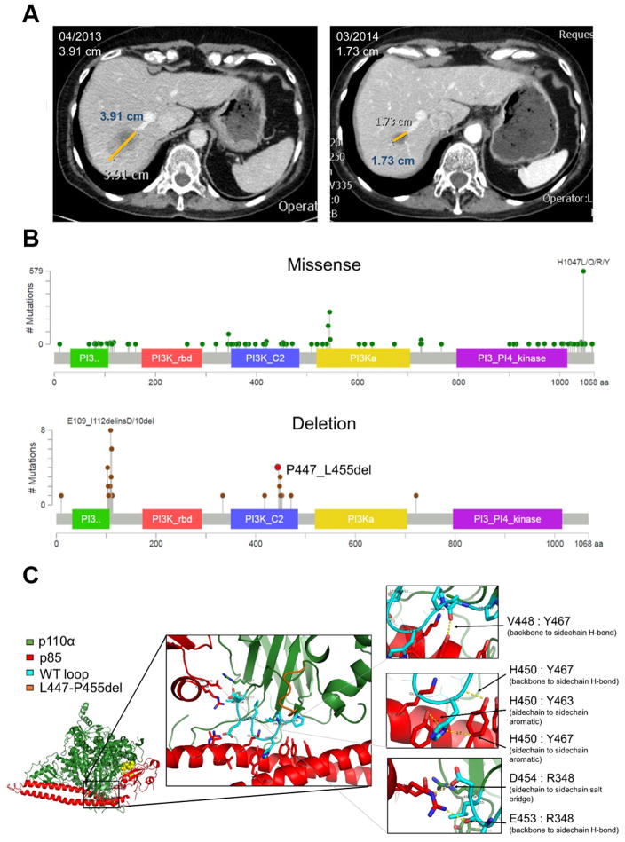
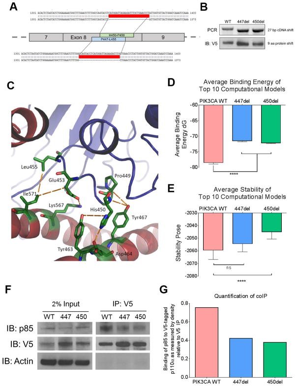
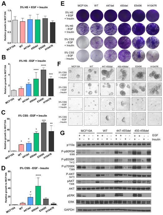
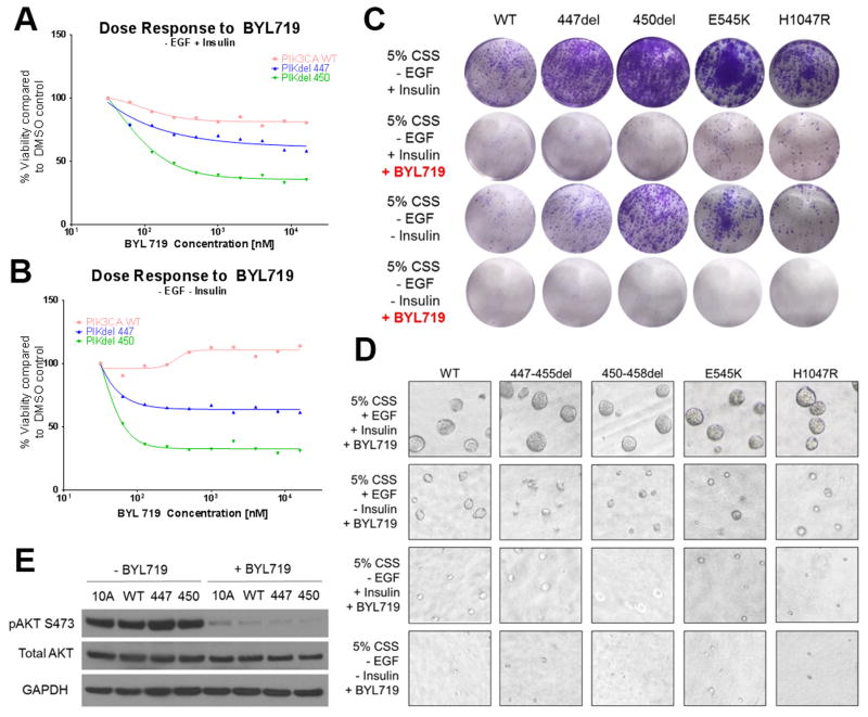
Purpose: We describe herein a novel P447_L455 deletion in the C2 domain of PIK3CA in a patient with an ER+ breast cancer with an excellent response to the PI3Kα inhibitor alpelisib. Although PIK3CA deletions are relatively rare, a significant portion of deletions cluster within amino acids 446-460 of the C2 domain, suggesting these residues are critical for p110α function.Experimental Design: A computational structural model of PIK3CAdelP447-L455 in complex with the p85 regulatory subunit and MCF10A cells expressing PIK3CAdelP447-L455 and PIK3CAH450_P458del were used to understand the phenotype of C2 domain deletions.Results: Computational modeling revealed specific favorable inter-residue contacts that would be lost as a result of the deletion, predicting a significant decrease in binding energy. Coimmunoprecipitation experiments showed reduced binding of the C2 deletion mutants with p85 compared with wild-type p110α. The MCF10A cells expressing PIK3CA C2 deletions exhibited growth factor-independent growth, an invasive phenotype, and higher phosphorylation of AKT, ERK, and S6 compared with parental MCF10A cells. All these changes were ablated by alpelisib treatment.Conclusions: C2 domain deletions in PIK3CA generate PI3K dependence and should be considered biomarkers of sensitivity to PI3K inhibitors. Clin Cancer Res; 24(6); 1426-35. ©2017 AACR.


Conflict of interest statement

Disclosure of potential conflicts of interest: R.N. and R.L. are employees of Guardant Health. J.H. and V.M. are employees of Foundation Medicine. LCC is a founder and holds equity in Agios Pharmaceuticals and Petra Pharmaceuticals, companies that are developing drugs for cancer therapy. The research in this paper does not involve drugs being developed at these companies

Figures

Fig. 1
Fig. 1. PIK3CA C2 deletions occur in breast cancer and cluster predominately at p85α binding sites
(A) Liver metastasis of patient with endocrine resistant ER+ breast cancer harboring the PIK3CAP447_L455del at baseline and 11 months after starting treatment with letrozole and alpelisib. (B) Lollipop plots of PIK3CA missense mutations (top) and deletions (bottom) from the cBioportal database (accessed 1/2017). (C) The heterodimeric structure of p110α (green) and p85α (red). Structural analysis determined the conformation of the PIK3CAWT p110α (cyan) and the interaction with p85α is altered by the deletion, PIK3CAP447-L455del (orange). Six major interaction points (arrows) are disrupted by the loss of the nine amino acids (right).
Fig. 2
Fig. 2. PIK3CA C2 deletions lead to disruption of p85α binding
(A) Schematic representation of targeting sites of deletions in PIK3CA, the patient deletion, PIK3CAP447_L455del (447del) and an additional deletion from cBioportal, PIK3CAH450_P458del (450del). The Gateway cloning system was used to design constructs with V5-tag and deleted sequences were confirmed (red). MCF10A cells were infected with lentivirus to stable express the PIK3CAWT and the two deletions which will be referred to the PIK3CA deletion panel. (B) PCR (top) and immunoblot of V5-tag expression (bottom) confirmation of the 27 base pair DNA and 9 amino acid protein shift, respectively. (C) p110α and p85α interacting residues within PIK3CAP447-L455del . Seven main disrupted interactions are listed in Table 1. (D) Average binding energy between p110α and p85α of the top 10 computational models. Both PIK3CAP447_L455del and PIK3CAH450_P458del exhibited a statistically significant decrease in binding energy (p<0.0001). (E) Average calculated stability of p110α in the top 10 computational models. Only PIK3CAH450_P458del exhibited a statistically significant decrease in stability (p<0.0001). (F) Co-immunoprecipitation of cell lysates from MCF10A cells stably expressing PIK3CAWT or the two C2 domain deletions. Protein (1 mg) was isolated and immunoprecipitated (IP) with 1 μg anti V5-Tag antibody. Lysates were then separated with SDS-PAGE and subjected to immunoblot analysis (IB) with a p85α antibody. (G) Immunoblots were quantified using ImageJ.
Fig. 3
Fig. 3. PIK3CA C2 deletions are activating in breast cancer
(A–D) Quantification of monolayer growth assay of stably transduced MCF10A cells in various media conditions lacking essential growth factors (*p > .05, ** p > .01, *** p > .001, **** p > .0001). Cells were seeded in triplicate in 12-well plates and (E) stained on day 8 with crystal violet. (F) Parental and stably transduced MCF10A cells were plated in 3D Matrigel in MCF10A media containing 5% charcoal stripped serum (CSS) ± EGF ± Insulin. Media and growth factors were changed every 3 days. Colonies (≥50 μM) were imaged on Day 12. Assay was carried out in duplicate wells in 3 separate experiments. (G) Cells were grown in MCF10A media containing 5% CSS and ± EGF ± Insulin for 24 h. Cell lysates were subjected to immunoblot analyses with the indicated antibodies.
Fig. 4
Fig. 4. PIK3CA C2 deletions are sensitive to PI3Kα inhibitor alpelisib
(A) Alpelisib dose response curve in the absence of EGF. The indicated MCF10A transfectants were seeded onto 96-well plates in triplicate and treated with increasing concentrations of alpelisib for 7 days. Plates were stained with crystal violet and resuspended in 1% sodium dodecyl sulfate (SDS) and read on a Promega GloMax Microplate reader. (B) Alpelisib dose response curve in the absence of EGF and insulin. (C) Growth assay with the PIK3CA deletion panel in the absence of EGF ± insulin ± 1μM alpelisib. Cells were seeded in triplicate in 12-well plates and stained on day 8 with crystal violet. Experiment was repeated 3 times. (D) Cells were plated in 3D Matrigel in MCF10A media containing 5% charcoal stripped serum (CSS) ± EGF ± insulin in the presence of 1 μM alpelisib. (E) Cells were grown in MCF10A media containing 5% CSS ± EGF ± insulin for 24 h in the presence of 1 μM alpelisib. Cell lysates were subjected to immunoblot analyses with the indicated antibodies.

Similar articles

Cited by

References

    1. Thorpe LM, Yuzugullu H, Zhao JJ. PI3K in cancer: divergent roles of isoforms, modes of activation and therapeutic targeting. Nat Rev Cancer. 2015;15(1):7–24. doi: 10.1038/nrc3860. - DOI - PMC - PubMed
    1. Samuels Y, Wang Z, Bardelli A, Silliman N, Ptak J, Szabo S, et al. High frequency of mutations of the PIK3CA gene in human cancers. Science. 2004;304(5670):554. doi: 10.1126/science.1096502. - DOI - PubMed
    1. Gymnopoulos M, Elsliger MA, Vogt PK. Rare cancer-specific mutations in PIK3CA show gain of function. Proc Natl Acad Sci U S A. 2007;104(13):5569–74. doi: 10.1073/pnas.0701005104. - DOI - PMC - PubMed
    1. Engelman JA. Targeting PI3K signalling in cancer: opportunities, challenges and limitations. Nat Rev Cancer. 2009;9(8):550–62. doi: 10.1038/nrc2664. - DOI - PubMed
    1. Huang TH, Huo L, Wang YN, Xia W, Wei Y, Chang SS, et al. Epidermal growth factor receptor potentiates MCM7-mediated DNA replication through tyrosine phosphorylation of Lyn kinase in human cancers. Cancer Cell. 2013;23(6):796–810. doi: 10.1016/j.ccr.2013.04.027. - DOI - PMC - PubMed

Publication types