. 2018 Jan 9;115(2):397-402.
doi: 10.1073/pnas.1713284115. Epub 2017 Dec 28.

Role of enhanced receptor engagement in the evolution of a pandemic acute hemorrhagic conjunctivitis virus

Affiliations

Role of enhanced receptor engagement in the evolution of a pandemic acute hemorrhagic conjunctivitis virus

Jim Baggen et al. Proc Natl Acad Sci U S A. .

Abstract

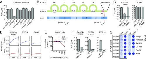
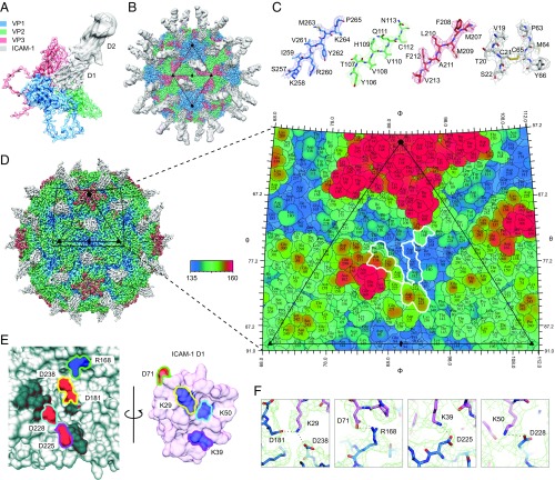
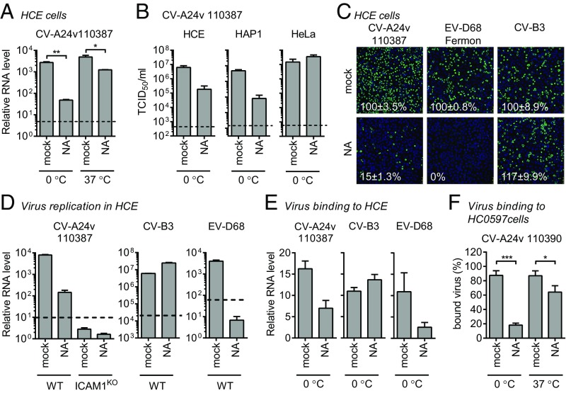
Acute hemorrhagic conjunctivitis (AHC) is a painful, contagious eye disease, with millions of cases in the last decades. Coxsackievirus A24 (CV-A24) was not originally associated with human disease, but in 1970 a pathogenic "variant" (CV-A24v) emerged, which is now the main cause of AHC. Initially, this variant circulated only in Southeast Asia, but it later spread worldwide, accounting for numerous AHC outbreaks and two pandemics. While both CV-A24 variant and nonvariant strains still circulate in humans, only variant strains cause AHC for reasons that are yet unknown. Since receptors are important determinants of viral tropism, we set out to map the CV-A24 receptor repertoire and establish whether changes in receptor preference have led to the increased pathogenicity and rapid spread of CV-A24v. Here, we identify ICAM-1 as an essential receptor for both AHC-causing and non-AHC strains. We provide a high-resolution cryo-EM structure of a virus-ICAM-1 complex, which revealed critical ICAM-1-binding residues. These data could help identify a possible conserved mode of receptor engagement among ICAM-1-binding enteroviruses and rhinoviruses. Moreover, we identify a single capsid substitution that has been adopted by all pandemic CV-A24v strains and we reveal that this adaptation enhances the capacity of CV-A24v to bind sialic acid. Our data elucidate the CV-A24v receptor repertoire and point to a role of enhanced receptor engagement in the adaptation to the eye, possibly enabling pandemic spread.

Keywords: ICAM-1; conjunctivitis; coxsackievirus A24v; receptor; sialic acid.


Conflict of interest statement

The authors declare no conflict of interest.

Figures

Fig. 1.
Fig. 1.
ICAM-1 is an essential CV-A24 receptor. (A) Yields of infectious virus after a single replication cycle. Sia-deficient HAP1 CMASKO cells were treated with antibodies against enterovirus receptors (ICAM-1, DAF, integrin α2, PVR, CAR, integrin αvβ3, and PSGL-1) or soluble receptor (VLDL-R) and infected at 37 °C with CV-A24v 110387. (B) Schematic representation of ICAM-1 showing Ig-like domains 1–5 (D1–D5) and the two sites targeted by CRISPR/Cas9 (gRNA 1 and 2). Knockout was accomplished by disrupting a 1,489-bp region of ICAM1 encoding the transmembrane domain (TM), either by excision of this region or by introducing a frameshift mutation. (C) HeLa-R19 ICAM-1KO transfected with plasmid encoding ICAM-1 cDNA were exposed to virus and yields of infectious virus were measured after a single replication cycle. (D) Biolayer interferometry analysis of virus binding to either ICAM-1 or negative control receptor LAMP-1. (E) Relative levels of CV-A24v 110390 bound to primary conjunctival cells with expanded lifespan (HC0597), in the presence of ICAM-1 D1D5 or negative control receptor CAR D1. (F) HeLa-R19 cells were infected and yields of infectious virus were determined after a single replication cycle. Dashed lines (A, C, and F) represent virus input levels (T = 0). Error bars (A, C, E, and F) represent the mean ± SEM of three to four biological replicates. (G) HeLa-R19 cells were infected with seven CV-A24v clinical isolates, two CV-A24 nonvariant strains and ICAM-1-dependent (CV-A21, RV-14) or CAR-dependent (CV-B3) control viruses, followed by crystal violet staining of surviving cells.
Fig. 2.
Fig. 2.
The structure of CV-A24v in complex with ICAM-1. (A) Enlarged tilted view of density denoted by a black triangle in B, containing the corresponding atomic model. ICAM-1 D1 binds in the “canyon” located at the quasi-threefold axis. (B) The cryo-EM reconstruction of CV-A24v in complex with ICAM-1 (D1D2) viewed down the icosahedral twofold axis. VP1-3 (3.6σ), D1 (1σ), and D2 (0.6σ). (C) Typical example of the cryo-EM electron density of VP1-3 and D1 domain. (D) Radially colored isosurface representation of CV-A24v in complex with D1 (gray) viewed down the icosahedral twofold axis (Left). The stereographic projection of the viral surface (Right), where the polar angles θ and φ represent latitude and longitude, respectively. Amino acids which interact with ICAM-1 D1 are located at the floor and wall of the canyon (blue) and are circled in white. The radial coloring key is shown in angstroms. (E) Surface representation of the EM-derived atomic model for the quasi-threefold axis of CV-A24v (gray) and ICAM-1 D1 (pink), with residues forming salt bridges labeled and colored in respect to their interacting partners. (Left) The surface of CV-A24v. (Right) The surface of ICAM-1 D1 that interacts with CV-A24v. Residues forming salt bridges are colored according to charge (red is negative, blue is positive) and capsid residues that hydrogen-bond with ICAM-1 are dark gray. (F) Overview of electrostatic interactions shown in E between ICAM-1 (pink) and CV-A24v (blue) with potential hydrogen bonds denoted as dotted black lines and EM density as a green mesh.
Fig. 3.
Fig. 3.
Sia is an attachment receptor supporting ICAM-1–mediated CV-A24v infection. (A) HCE cells were treated with a mixture of A. urefaciens and Vibrio cholerae NA (1:30), infected with CV-A24v 110387 on ice (0 °C) or at 37 °C, followed by qPCR analysis of viral RNA levels after a single replication cycle. (B) NA-treated cells were infected with CV-A24v 110387 and yields of infectious virus were determined after a single replication cycle. (C) NA-treated HCE cells were infected on ice with CV-A24v, Sia-dependent enterovirus D68 (EV-D68), and Sia-independent CV-B3, followed by staining of dsRNA (green) and nuclei (blue). Shown are representative confocal micrographs. Percentage values denote mean ± SEM of three technical replicates, normalized to mock. (D and E) HCE cells were treated with a mixture of A. urefaciens and V. cholerae NA (1:30) and infected on ice. After incubation with virus, viral RNA levels were determined by qPCR either directly (E) or after a single replication cycle (D). (F) Levels of radioactively labeled CV-A24v 110390 bound to NA-treated (V. cholerae 10 mU/mL) primary conjunctival cells with expanded lifespan (HC0597) on ice (0 °C) or at 37 °C. Dashed lines (A, B, and D) represent virus input levels (T = 0). Error bars represent the mean ± SEM of three (A, B, D, and E) or eight (F) biological replicates. P values (A and F) were calculated by an unpaired two-sided t test; *P < 0.05; **P < 0.01; ***P < 0.001.
Fig. 4.
Fig. 4.
Pandemic CV-A24v strains have acquired a capsid residue that enhances Sia binding. (A) NA-treated HCE cells were infected on ice, followed by staining of infected cells with a dsRNA antibody (green) and nuclei with DAPI (blue). Percentage values denote mean ± SEM of three technical replicates, normalized to mock. (B) Amino acid sequence alignment of tested CV-A24 strains, with the residue unique for nonvariant strains highlighted in red. (C) Binding site for Sia (orange) in CV-A24v 110390 (PDB ID code 4Q4X), showing that the 5-N-acyl group of Sia is stabilized by two hydrogen bonds (dashed lines). Two adjacent VP1 proteins are colored gray and blue, respectively. Red spheres represent water molecules. Oxygen and nitrogen atoms are colored red and blue, respectively. (D) Geographic origins of non-AHC- (red) and AHC-causing (purple, light blue, blue) CV-A24 strains of which complete VP1 sequences are available, with circle sizes proportional to the number of isolates. (E) Phylogenetic tree of CV-A24 isolates, showing on the right side the frequencies of amino acids in the Sia-binding site among the indicated number of strains. Frequency plots were generated with WebLogo. Residues at VP1 position 250 are highlighted in gray. (F) HCE cells were treated with NA and infected on ice with CV-A24v 110390 or the VP1 Y250F mutant, followed by staining of dsRNA (green) and nuclei (blue). Quantifications shown at the bottom denote mean ± SEM of four technical and two biological replicates. Images shown in A and F are representative confocal micrographs. (G) HCE cells were treated with a mixture of A. urefaciens and V. cholerae NA (1:30) and incubated with virus on ice, followed by qPCR analysis of bound virus. Error bars represent the mean ± SEM of three biological replicates. P values (F and G) were calculated by an unpaired two-sided t test; not significant (ns), P ≥ 0.05; **P < 0.01; ****P < 0.0001.

References

    1. Ghazali O, et al. An outbreak of acute haemorrhagic conjunctivitis in Melaka, Malaysia. Singapore Med J. 2003;44:511–516. - PubMed
    1. Kurokawa M, Rai SK, Ono K, Gurung R, Ruit S. Viral investigation of acute hemorrhagic conjunctivitis outbreak (2003) in Nepal using molecular methods. Southeast Asian J Trop Med Public Health. 2006;37:904–910. - PubMed
    1. Oh MD, et al. Acute hemorrhagic conjunctivitis caused by coxsackievirus A24 variant, South Korea, 2002. Emerg Infect Dis. 2003;9:1010–1012. - PMC - PubMed
    1. Measroch V, Gear J, Faerber GI. Studies in poliomyelitis; the isolation of a coxsackie-like virus from the faeces of apparently healthy bantu infants. S Afr Med J. 1951;25:421–424. - PubMed
    1. Kono R, Sasagawa A, Ishii K, Sugiura S, Ochi M. Pandemic of new type of conjunctivitis. Lancet. 1972;1:1191–1194. - PubMed

Publication types

MeSH terms