

Weight loss in the healthy elderly might be a non-cognitive sign of preclinical Alzheimer's disease
- PMID: 29285207
- PMCID: PMC5739594
- DOI: 10.18632/oncotarget.22218
Weight loss in the healthy elderly might be a non-cognitive sign of preclinical Alzheimer's disease
Abstract
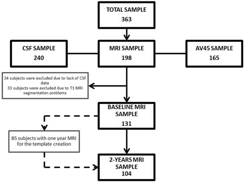
Weight loss has been proposed as a sign of pre-clinical Alzheimer Disease (AD). To test this hypothesis, we have evaluated the association between longitudinal changes in weight trajectories, cognitive performance, AD biomarker profiles and brain structure in 363 healthy controls from the Alzheimer´s Disease Neuroimaging Initiative (mean follow-up 50.5±30.5 months). Subjects were classified according to body weight trajectory into a weight loss group (WLG; relative weight loss ≥ 5%) and a non-weight loss group (non-WLG; relative weight loss < 5%). Linear mixed effects models were used to estimate the effect of body weight changes on ADAS-Cognitive score across time. Baseline CSF tau/AΔ42 ratio and AV45 PET uptake were compared between WLG and non-WLG by analysis of covariance. Atrophy maps were compared between groups at baseline and longitudinally at a 2-year follow-up using Freesurfer. WLG showed increased baseline levels of cerebrospinal fluid tau/AΔ42 ratio, increased PET amyloid uptake and diminished cortical thickness at baseline. WLG also showed faster cognitive decline and faster longitudinal atrophy. Our data support weight loss as a non-cognitive manifestation of pre-clinical AD.
Keywords: Gerotarget; PET amyloid; cerebrospinal fluid Alzheimer’s disease biomarkers; magnetic resonance imaging; pre-clinical Alzheimer’s disease; weight loss.
Conflict of interest statement
CONFLICTS OF INTEREST All authors report no biomedical financial interests or potential conflicts of interest related to this work.
Figures


References
-
- Ingram DD, Mussolino ME. Weight loss from maximum body weight and mortality: the Third National Health and Nutrition Examination Survey Linked Mortality File. Int J Obes. 2010;34:1044–50. - PubMed
-
- Pizzato S, Sergi G, Bolzetta F, De Rui M, De Ronch I, Carraro S, Berton L, Orr E, Imoscopi A, Perissinotto E, Coin A, Manzato E, Veronese N. Effect of weight loss on mortality in overweight and obese nursing home residents during a 5-year follow-up. Eur J Clin Nutr. 2015;69:1113–18. - PubMed
-
- Gillette-Guyonnet S, Nourhashemi F, Andrieu S, de Glisezinski I, Ousset PJ, Riviere D, Albarede JL, Vellas B. Weight loss in Alzheimer disease. Am J Clin Nutr. 2000;71:637S–42S. - PubMed
-
- White H, Pieper C, Schmader K. The association of weight change in Alzheimer's disease with severity of disease and mortality: a longitudinal analysis. J Am Geriatr Soc. 1998;46:1223–27. - PubMed
Grants and funding
LinkOut - more resources
Full Text Sources
Other Literature Sources
