. 2017 Nov 6;8(62):105584-105595.
doi: 10.18632/oncotarget.22331. eCollection 2017 Dec 1.

Lipopolysaccharide-coated CuS nanoparticles promoted anti-cancer and anti-metastatic effect by immuno-photothermal therapy

Affiliations

Lipopolysaccharide-coated CuS nanoparticles promoted anti-cancer and anti-metastatic effect by immuno-photothermal therapy

Bian Jang et al. Oncotarget. .

Abstract

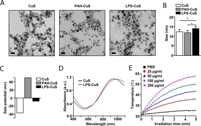
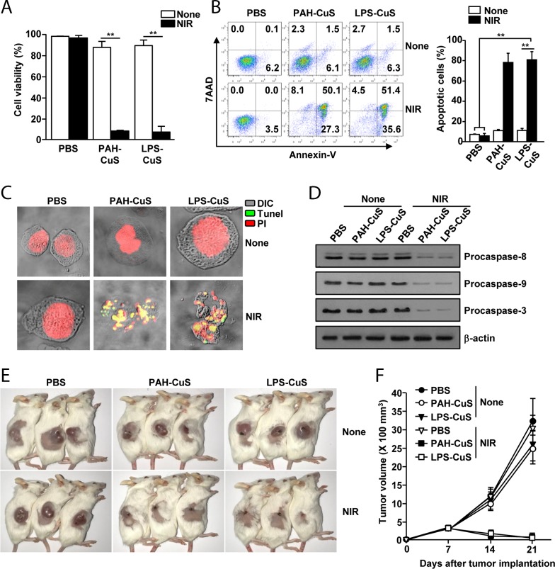
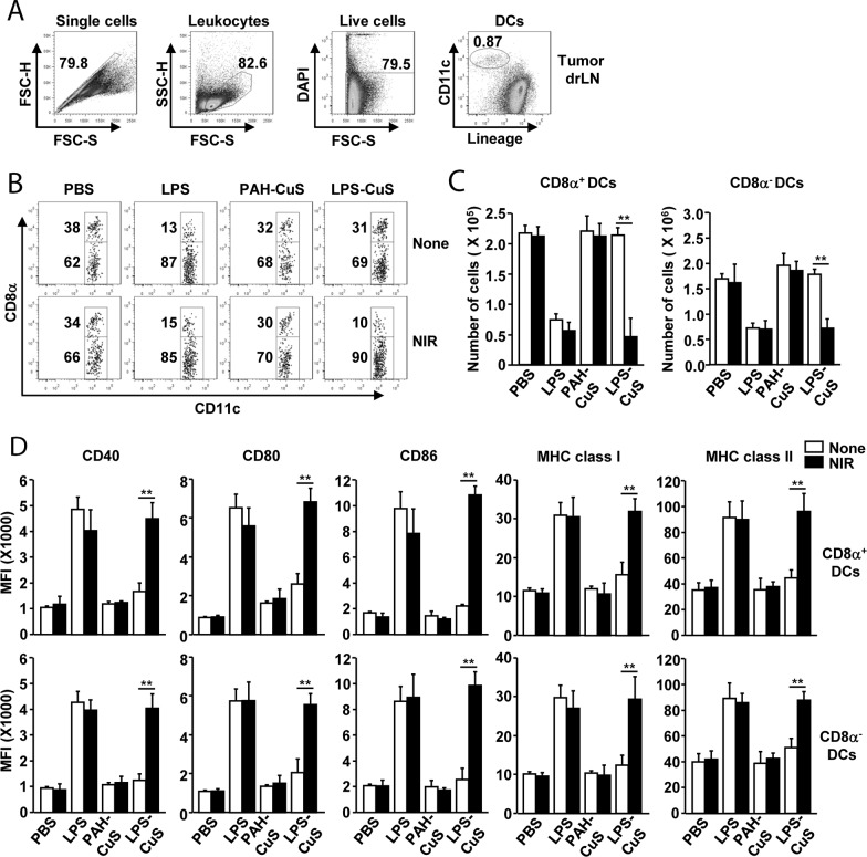
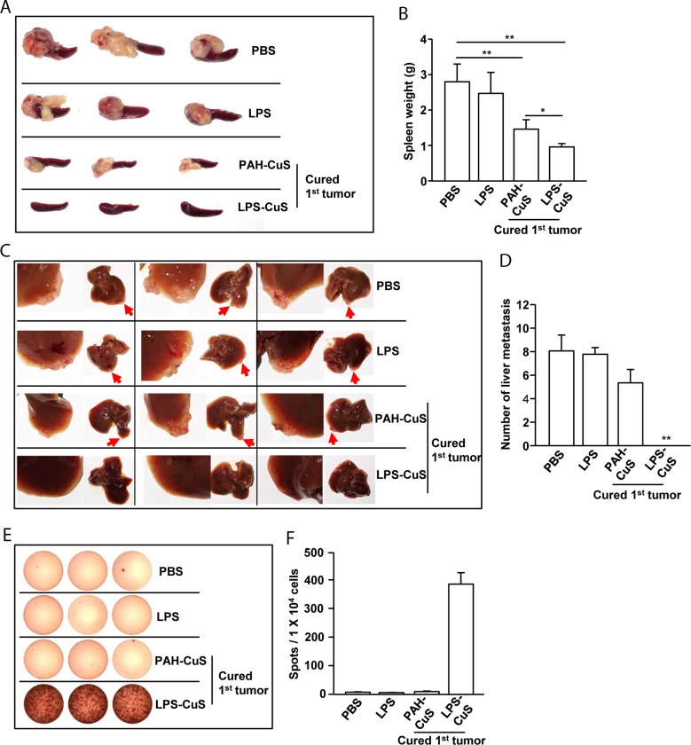
To meet the ultimate goal of cancer therapy, which is treating not only the primary tumor but also preventing metastatic cancer, the concept of combining immunotherapy with photothermal therapy (PTT) is gaining great interest. Here, we studied the new material, lipopolysaccharide (LPS) coated copper sulfide nanoparticles (LPS-CuS), for the immuno-photothermal therapy. We evaluated the effect of LPS-CuS for induction of apoptosis of CT26 cells and activation of dendritic cells. Moreover, the LPS-CuS and laser irradiation was examined anti-metastasis effect by liver metastasis model mouse in vivo. Through PTT, LPS-CuS induced elimination of CT26 tumor in BALB/c mice, which produced cancer antigens. In addition, released LPS and cancer antigen by PTT promoted dendritic cell activation in tumor draining lymph node (drLN), and consequently, enhanced the tumor antigen-specific immune responses. Finally, the primary tumor cured mice by LPS-CuS-mediated PTT completely resisted secondary tumor injection in the spleen and also prevented liver metastasis. Our results demonstrated the potential usage of LPS-CuS for the immuno-photothermal therapy against various types of cancer by showing the clear elimination of primary colon carcinoma with complete prevention of spleen and liver metastasis.

Keywords: anti-tumor; copper sulfide nanoparticles; immunotherapy; lipopolysaccharide; photothermal therapy.


Conflict of interest statement

CONFLICTS OF INTEREST Competing financial interests: The authors declare no competing financial interest.

Figures

Figure 1
Figure 1. Schematic diagrams
Structural illustration of LPS-CuS.
Figure 2
Figure 2. Characterization of F-CuS
(A) FE-TEM images of CuS, PAH-CuS, and LPS-CuS. (Scale bars: 20 nm). (B) TEM corresponding size distribution of each coated nanoparticles. (C) Zeta potential values of each NPs. (D) UV-vis absorption of CuS (black) and LPS-CuS (red). (E) Photothermal heating curves of different concentrations of LPS-CuS dissolved in water, irradiated for 5 min with an 808-nm laser at a power density of 1 W/cm2.
Figure 3
Figure 3. LPS-CuS with laser irradiation promoted anti-cancer effect against CT26 cells in vitro and in vivo
CT26 cells were incubated with PBS, PAH-CuS, and LPS-CuS for 2 h, and the cells were treated with or without laser irradiation at 1 W/cm2 for 5 min and cultured for 24 h. (A) Cell viability of CT26 was measured by MTT assay; ** p < 0.01. (B) Apoptosis were analyzed by annexin-V and 7AAD staining (left panel). Mean percentages of apoptotic cells were shown (right panel), ** p < 0.01. (C) Nuclear degradation was analyzed by Tunel assay. (D) The expression levels of procaspase-8, -9, and -3 were measured by western blotting analysis. β-actin were used as a loading control. (E-F) BLAB/c mice were subcutaneously inoculated with 1 × 10⁶ CT26 cells. The mice were intratumorally injected with PBS, PAH-CuS, and LPS-CuS for 2 h 7 days after tumor injection and were treated with or without laser irradiation for 5 min. (E) The sizes of tumor mass on day 21 after tumor injection are shown. (F) CT26 tumor growth curves for the mice. Data are from the analyses of six individual mice (three mice per experiment, for a total of two independent experiments).
Figure 4
Figure 4. LPS-CuS with laser irradiation promoted DC activation in tumor draining lymph node (drLN)
CT26 tumor-bearing mice were intratumorally injected with PBS, LPS, PAH-CuS, and LPS-CuS for 2 h, and treated with or without laser irradiation for 5 min. Tumor drLN were harvested 24 h after treatment. (A) Definition of DC population in tumor drLN. Lineage markers included CD3, Thy1.1, B220, Gr-1, CD49b, and TER-119. (B) Lineage-CD11c+ DCs were further divided as CD8α+ and CD8α DCs. (C) Mean number of CD8α+ and CD8αDCs in tumor drLN. (D) Mean fluorescence intensity (MFI) of co-stimulatory molecules and MHC class I and II in gated CD8α+ and CD8α DCs in tumor drLN were analyzed using flow cytometry. All data are representative of or the average of analyses of six independent samples (i.e., three samples per experiment, two independent experiments).
Figure 5
Figure 5. LPS-CuS treatment with laser irradiation prevented liver metastasis of CT26 cells
On day 21 of primary tumor challenge, PAH-CuS and LPS-CuS treatment with laser irradiated mice were intrasplenically injected with secondary CT26 cells. PBS and LPS-treated mice were also intrasplenically injected with CT26 cells. (A) The size of tumor mass in the spleen on day 14 after secondary CT26 injection. (B) Mean weights of spleens. (C) Liver metastasis of CT26 tumor was measured. Red arrows indicated tumor mass in the liver. (D) The mean of the absolute number of CT26 metastasis in the livers. (E) Splenocytes were stimulated with CT26 self-antigen for 24 h. IFN-γ production were analyzed by ELISPOT analysis. (F) The mean number of spots was shown. Data are representative of analyses of six independent samples (i.e., three mice per experiment, two independent experiments).

References

    1. Chaffer CL, Weinberg RA. A perspective on cancer cell metastasis. Science. 2011;331:1559–64. https://doi.org/10.1126/science.1203543 - DOI - PubMed
    1. Britten CM, Singh-Jasuja H, Flamion B, Hoos A, Huber C, Kallen KJ, Khleif SN, Kreiter S, Nielsen M, Rammensee HG, Sahin U, Hinz T, Kalinke U. The regulatory landscape for actively personalized cancer immunotherapies. Nat Biotechnol. 2013;31:880–2. https://doi.org/10.1038/nbt.2708 - DOI - PubMed
    1. Oldham RK, Dillman RO. Monoclonal antibodies in cancer therapy: 25 years of progress. J Clin Oncol. 2008;26:1774–7. https://doi.org/10.1200/JCO.2007.15.7438 - DOI - PubMed
    1. Gattinoni L, Powell DJ, Jr, Rosenberg SA, Restifo NP. Adoptive immunotherapy for cancer: building on success. Nat Rev Immunol. 2006;6:383–93. https://doi.org/10.1038/nri1842 - DOI - PMC - PubMed
    1. Gubin MM, Zhang X, Schuster H, Caron E, Ward JP, Noguchi T, Ivanova Y, Hundal J, Arthur CD, Krebber WJ, Mulder GE, Toebes M, Vesely MD, et al. Checkpoint blockade cancer immunotherapy targets tumour-specific mutant antigens. Nature. 2014;515:577–81. https://doi.org/10.1038/nature13988 - DOI - PMC - PubMed

LinkOut - more resources