. 2017 Nov 18;8(62):105923-105935.
doi: 10.18632/oncotarget.22510. eCollection 2017 Dec 1.

Insulin receptor substrate-4 interacts with ubiquitin-specific protease 18 to activate the Jak/STAT signaling pathway

Affiliations

Insulin receptor substrate-4 interacts with ubiquitin-specific protease 18 to activate the Jak/STAT signaling pathway

Baihai Jiao et al. Oncotarget. .

Abstract

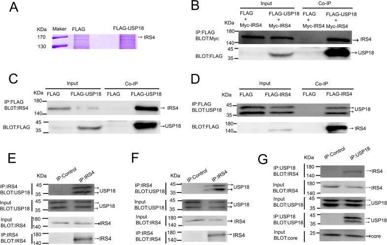
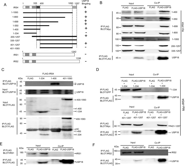
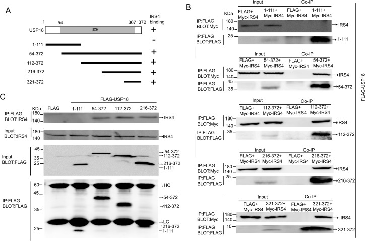
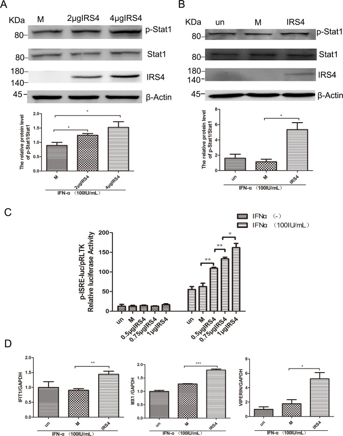
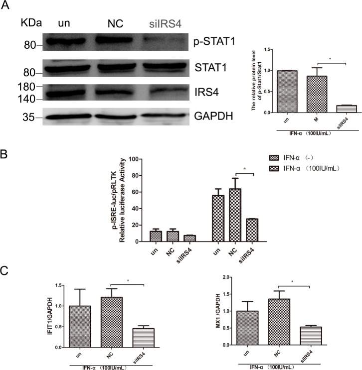
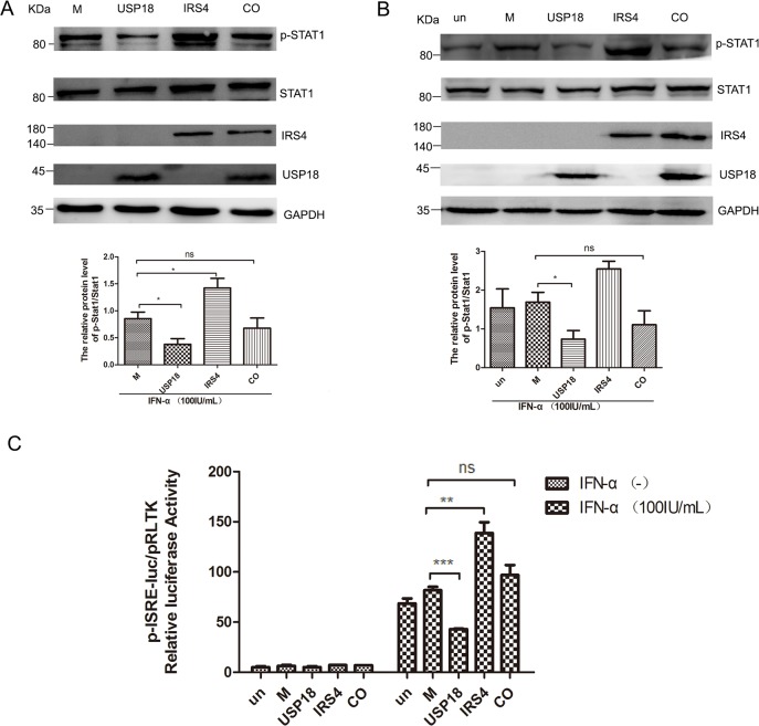
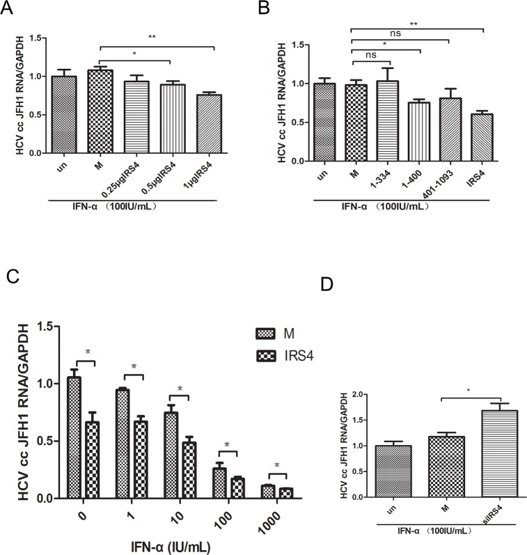
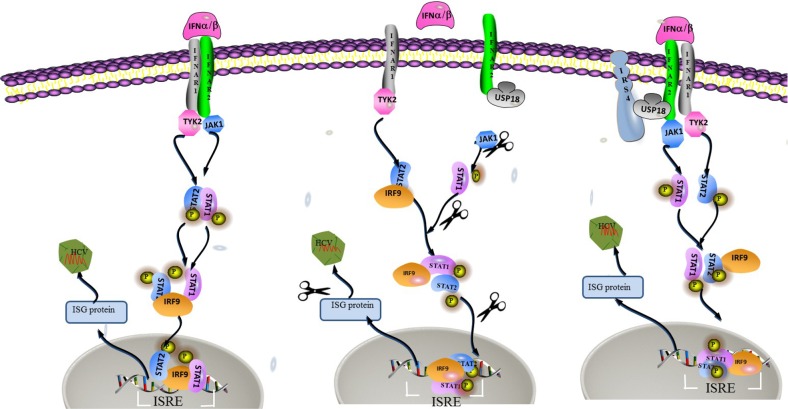
Ubiquitin-specific protease 18 (USP18) as a negative regulator of the Jak/STAT signaling pathway plays an important role in the host innate immune response. USP18 has been shown to bind to the type I interferon receptor subunit 2 (IFNAR2) to down-regulate the Jak/STAT signaling. In this study, we showed that insulin receptor substrate (IRS)-4 functioned as a novel USP18-binding protein. Co-precipitation assays revealed that two regions (amino acids 335-400 and 1094-1257) of IRS4 were related to bind to the C- terminal region of USP18. IRS4 binding to USP18 diminished the inhibitory effect of USP18 on Jak/STAT signaling. IRS4 over-expression enhanced while IRS4 knock-down suppressed the Jak/STAT signaling in the presence of IFN-a stimulation. As such, IRS4 increased IFN-a-mediated anti-HCV activity. Mechanistically, IRS4 promoted the IFN-a-induced Jak/STAT signaling by interact with USP18. These results suggested that IRS4 binds to USP18 to diminish the blunting effect of USP18 on IFN-a-induced Jak/STAT signaling. Our findings indicated that IRS4 is a novel USP18-binding protein that can be used to boost the host innate immunity to control HCV, and potentially other viruses that are sensitive to IFN-a.

Keywords: HCV; IRS4; Jak/STAT signaling pathway; USP18.


Conflict of interest statement

CONFLICTS OF INTEREST The authors disclose no conflicts of interest.

Figures

Figure 1
Figure 1. USP18 interacted with IRS4
(A) identification of USP18-binding proteins. 293T cell lines stably expressing Flag or Flag-USP18 were established. Cell protein lysates were precipitated with anti-Flag M2 affinity gel. The immunoprecipitated protein complexes were separated by SDS–PAGE, stained using Coomassie R-350 and analyzed by mass spectrometric analysis. (B) IRS4 interacted with USP18. 293T cells were co-transfected with Myc-IRS4 and Flag-USP18 or Flag empty plasmid. Cell lysates were immunoprecipitated (IP) with an anti-Flag antibody (incubated with anti-Flag M2 Affinity Gel) and analyzed by immunoblotting with anti-Myc (upper) and anti-Flag antibodies (lower). (C) Co-IP assay of the interaction between USP18 and endogenous IRS4. 293T cells were transfected with Flag or Flag-USP18. Cell protein lysates were immunoprecipitated with an anti-Flag antibody followed by immunoblotting with anti-IRS4(upper) and anti-Flag antibodies (lower). (D) Co-IP assay of the interaction between IRS4 and endogenous USP18. 293T cells were transfected with Flag or Flag-IRS4. Cell protein lysates were immunoprecipitated with an anti-Flag antibody followed by immunoblotting with anti-USP18(upper) and anti-Flag antibodies (lower). (E, F) IRS4 interacts with USP18 endogenously. 293T cells(E) and Huh7.5.1 cells(F) lysates were immunoprecipitated with an anti-IRS4 antibody or control IgG and analyzed by immunoblotting with anti-USP18 and anti-IRS4 antibodies. (G) IRS4 interacts with USP18 endogenously in JFH1-infected Huh7.5.1 cells. JFH1-infected Huh7.5.1 cells lysates were immunoprecipitated with an anti-IRS4 antibody or control IgG and analyzed by immunoblotting with anti-USP18, anti-IRS4 and anti-core antibodies.
Figure 2
Figure 2. Mapping of USP18-binding regions of IRS4
(A) Schematic depiction of human IRS4 and its deletion mutants used in this study. PH and PTB indicate the pleckstrin homology (PH) domain and phosphotyrosine-binding (PTB) domain, respectively. Summary of the binding domains of IRS4 with USP18 was listed on the right. A major USP18-binding region (amino acids 335–400 and 1094-1257) is indicated. (B–D) co-immunoprecipitation assays. (B) 293T cells co-expressing Flag-USP18 and Myc-IRS4 mutant's proteins were lysed and immunoprecipitated with an anti-Flag antibody. Immunoprecipitates (IP) and cell protein lysates (Input) were analyzed by immunoblotting with anti-Myc and anti-Flag antibodies. The figure shows only the bait protein of 1-400. (C) Co-IP assay of the interaction between three IRS4 mutants and endogenous USP18. Cell protein lysates were then immunoprecipitated with an anti-Flag antibody followed by immunoblotting with anti-USP18 and anti-Flag antibodies. HC, heavy chain. (D) 293T cells co-expressing Flag-USP18 and YFP-IRS4 mutant's proteins was lysed and immunoprecipitated with an anti-Flag antibody. Immunoprecipitates (IP) and cell protein lysates (Input) were analyzed by immunoblotting with anti-GFP and anti-Flag antibodies. (E, F) Co-IP assay of the interaction between USP18 and endogenous IRS1 and IRS2. Cell lysates were immunoprecipitated with an anti-Flag antibody followed by immunoblotting with anti-IRS1 or -IRS2(upper) and anti-Flag antibodies (lower).
Figure 3
Figure 3. Mapping of IRS4-binding regions of USP18
(A) Schematic structure of human USP18 and its deletion mutants used in this study, UCH indicate the UCH (ubiquitin C-terminal hydrolase) domains. The IRS4 binding domains is indicated in the right column. (B) Co-IP assay of the interaction between USP18 mutants and Myc-IRS4. 293T cells co-expressing Flag-USP18 mutants and Myc-IRS4 were lysed and immunoprecipitated with an anti-Flag antibody. Immunoprecipitates and cell lysates were analyzed by immunoblotting with anti-Myc or anti-Flag antibody. (C) Co-IP assay of the interaction between USP18 mutants and endogenous IRS4. Cell protein lysates were then immunoprecipitated with an anti-Flag antibody followed by immunoblotting with anti-IRS4(upper) or anti-Flag antibody (lower). HC, heavy chain; LC, light chain.
Figure 4
Figure 4. Overexpression of IRS4 enhanced IFN-a-induced activation of Jak/STAT signalling
(A, B) Overexpression of IRS4 enhanced IFN-a stimulated p-STAT1 level. 293T cells (A) or Huh-7.5.1 cells (B) were transfected with Flag (M, mock control), Flag-IRS4 or untreated(un), the cells were treated with 100 IU/mL IFN-α for 30 mins at 48 hours after transfection. Protein lysates were harvested, separated by SDS-PAGE, and probed for STAT1-phospho701. Blots then were stripped and probed for total STAT1 expression. The ratio of p-STAT1 to total STAT1 from 3 independent experiments was quantified (bottom). (C) Overexpression of IRS4 promoted IFN-a-stimulated ISRE activity. Huh7.5.1 cells were co-transfected with Flag-IRS4 plasmid or Flag empty plasmid and pISRE-luc (expressing firefly luciferase) and pRL-TK (expressing Renilla luciferase) as described in Materials and Methods. (D) Overexpression of IRS4 promoted IFN-a-induced ISG expression. Expression levels of IFIT1/VIPERIN/MX1 were examined by real time PCR in Huh7.5.1 cells transfected with IRS4 plasmid, empty plasmid (M, mock control) or untreated (un) 24 hours after transfection and then treated with 100IU/mL IFN-α for 24 hours. Error bars indicated mean±SD, *P<0.05, **P<0.01, ***P<0.001.
Figure 5
Figure 5. IRS4 knock-down suppressed the IFN-a-induced activation of Jak/STAT signaling
(A) IRS4 knock-down inhibited IFN stimulated p-STAT1 level. Huh-7.5.1 cells were transfected with siIRS4, negative control (NC) or untreated (un), the cells were treated with 100 IU/mL IFN-α for 30 min at 48 hours after transfection. Protein lysates were harvested, separated electrophoretically, and probed for STAT1-phospho701. Blots then were stripped and probed for total STAT1 expression. The ratio of p-STAT1 to total STAT1 from 3 independent experiments was quantified (right). (B) IRS4 knock-down inhibited IFN stimulated ISRE activity. Huh7.5.1 cells were co-transfected siRNA and negative control with pISRE-luc (expressing firefly luciferase) and pRL-TK (expressing Renilla luciferase) as described in Materials and Methods. 24 hours later, cells were treated with 100 IU/mL IFN-α for 24 hours before the cells were lysed for dual luciferase reporter gene assay. (C) IRS4 knock-down inhibited IFNa-induced ISG expression. Expression levels of IFIT1/MX1 were examined by real time PCR in Huh7.5.1 cells transfected with siRNA and negative control (NC) or untreated (un), 24 hours after transfection and then treated with 100IU/mL IFN-α for 24 hours. Error bars indicated mean±SD, *P<0.05.
Figure 6
Figure 6. IRS4 promoted IFN-a-induced Jak/STAT signaling pathway through interact with USP18
IRS4 diminished the inhibitory effects of USP18 on Jak/STAT signaling pathway (A-C). 293T cells (A) and Huh7.5.1 cells (B) were transfected with single empty plasmid (M, mock control), Flag-USP18 and Myc-IRS4, or co-transfected with USP18 and IRS4. The cells were treated with 100IU/mL IFN-α for 30 mins at 48 hours after transfection. Protein lysates were harvested, separated by SDS-PAGE, and probed for STAT1-phospho701 and total STAT1 expression. The ratio of p-STAT1 to total STAT1 from 3 independent experiments was quantified (bottom). (C) USP18 expression Flag-tagged plasmid (M, mock control), IRS4 expression Myc-tagged plasmid were co-transfected together with plasmids pISRE-luc and pRL-TK into Huh7.5.1 cells as described in Materials and Methods. 24 hours later, cells were treated with 100IU/mL IFN-α for 24 hours before the cells were lysed for dual luciferase reporter gene assay for ISRE activity. Error bars indicated mean±SD, *P<0.05, **P<0.01, ***P<0.001.
Figure 7
Figure 7. IRS4 regulated HCV replication in JFH1 infected Huh7.5.1 cells
Different doses of IRS4 plasmid, Flag-tag empty plasmid (M, mock control) or nothing (un) were transfected into Huh7.5.1 cells infected with HCV JFH1. 24 hours later the cells were treated with 100IU/mL IFN-α (A) for 24 hours and the relative expression levels of HCV RNA were examined by real time PCR. (B) Two mutants IRS4 (1-334) and IRS4 (401–1093) that do not bind to USP18 have no effect on interferon anti-HCV activity. Flag empty vector (M, mock control) or IRS4 mutant 1-334 or 401-1093 were transfected into Huh7.5.1 cells infected with HCV JFH1. 24 hours later the cells were treated with 100 IU/mL IFN-α (A) for 24 hours and the relative expression levels of HCV RNA were examined by real time PCR. (C) Dosage effects of IFN-α (0, 1, 10, 100, 1000 IU/mL) on HCV replication in IRS4-overexpressed Huh7.5.1 cells infected with HCV JFH1. Relative expression levels of HCV RNA were examined by real time PCR. The values are displayed as the expression level of JFH1 HCV relative to Flag-tag empty vector (mock). Error bars indicated mean±SD, *P<0.05, **P<0.01. (D) IRS4 knock-down inhibited IFN-α induced anti-HCV activity. JFH1 infected Huh7.5.1 cells were transfected with siIRS4, negative control (NC) or untreated (un), 24 hours later the cells were treated with 100 IU/mL IFN-α for 24 hours and the relative expression levels of HCV RNA were examined by real time PCR.
Figure 8
Figure 8. A hypothetical model of IRS4/USP18 interaction and its role in IFN antiviral function
USP18 binding to IFNAR2 in vivo blocks the interaction between Jak and the IFN receptor, thereby reduces the phosphorylation of the receptor and STATs and suppresses signal pathway. The interaction between IRS4 and USP18 decreased the inhibitory effect of USP18 on Jak/STAT signaling pathway. Therefore, IRS4 enhanced IFN-α induced activation of the Jak/STAT signaling pathway as shown by the increased levels of p-STAT1 and enhanced ISRE activity and increased induction of ISGs.

Similar articles

Cited by

References

    1. Takaoka A, Yanai H. Interferon signalling network in innate defence. Cell Microbiol. 2006;8:907–922. - PubMed
    1. Hwang SY, Hertzog PJ, Holland KA, Sumarsono SH, Tymms MJ, Hamilton JA, Whitty G, Bertoncello I, Kola I. A null mutation in the gene encoding a type I interferon receptor component eliminates antiproliferative and antiviral responses to interferons alpha and beta and alters macrophage responses. Proc Natl Acad Sci U S A. 1995;92:11284–11288. - PMC - PubMed
    1. Muller U, Steinhoff U, Reis LF, Hemmi S, Pavlovic J, Zinkernagel RM, Aguet M. Functional role of type I and type II interferons in antiviral defense. Science. 1994;264:1918–1921. - PubMed
    1. Swann JB, Hayakawa Y, Zerafa N, Sheehan KC, Scott B, Schreiber RD, Hertzog P, Smyth MJ. Type I IFN contributes to NK cell homeostasis, activation, and antitumor function. J Immunol. 2007;178:7540–7549. - PubMed
    1. de Veer MJ, Holko M, Frevel M, Walker E, Der S Paranjape JM, Silverman RH, Williams BRG. Functional classification of interferon-stimulated genes identified using microarrays. J Leukoc Biol. 2001;69:912. - PubMed