

Role of Cone Beam Computed Tomography in Diagnosis and Treatment Planning in Dentistry: An Update
- PMID: 29285467
- PMCID: PMC5730974
- DOI: 10.4103/jispcd.JISPCD_516_16
Role of Cone Beam Computed Tomography in Diagnosis and Treatment Planning in Dentistry: An Update
Abstract
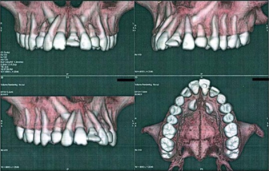
Accurate diagnosis and treatment planning are the backbone of any medical therapy; for this reason, cone beam computed tomography (CBCT) was introduced and has been widely used. CBCT technology provides a three-dimensional image viewing, enabling exact location and extent of lesions or any anatomical region. For the very same reason, CBCT can not only be used for surgical fields but also for fields such as endodontics, prosthodontics, and orthodontics for appropriate treatment planning and effective dental care. The aim and clinical significance of this review are to update dental clinicians on the CBCT applications in each dental specialty for an appropriate diagnosis and more predictable treatment.
Keywords: Anatomical variation; X-ray; cone beam computed tomography; dental technology; pathology; radiology; three-dimensional.
Conflict of interest statement
There are no conflicts of interest.
Figures
References
-
- Filler AG. The history, development, and impact of computed imaging in neurological diagnosis and neurosurgery: CT, MRI and DTI. Nat Proc. 2010;7:1–85.
-
- Orentlicher G, Goldsmith D, Abboud M. Computer-guided planning and placement of dental implants. Atlas Oral Maxillofac Surg Clin North Am. 2012;20:53–79. - PubMed
Publication types
LinkOut - more resources
Full Text Sources
Other Literature Sources
