. 2017 Oct;4(4):044504.
doi: 10.1117/1.JMI.4.4.044504. Epub 2017 Dec 14.

Context-aware stacked convolutional neural networks for classification of breast carcinomas in whole-slide histopathology images

Affiliations

Context-aware stacked convolutional neural networks for classification of breast carcinomas in whole-slide histopathology images

Babak Ehteshami Bejnordi et al. J Med Imaging (Bellingham). 2017 Oct.

Abstract

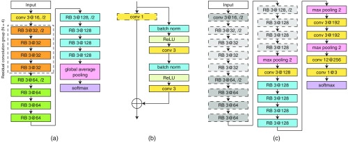
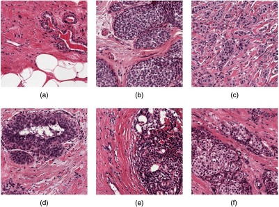
Currently, histopathological tissue examination by a pathologist represents the gold standard for breast lesion diagnostics. Automated classification of histopathological whole-slide images (WSIs) is challenging owing to the wide range of appearances of benign lesions and the visual similarity of ductal carcinoma in-situ (DCIS) to invasive lesions at the cellular level. Consequently, analysis of tissue at high resolutions with a large contextual area is necessary. We present context-aware stacked convolutional neural networks (CNN) for classification of breast WSIs into normal/benign, DCIS, and invasive ductal carcinoma (IDC). We first train a CNN using high pixel resolution to capture cellular level information. The feature responses generated by this model are then fed as input to a second CNN, stacked on top of the first. Training of this stacked architecture with large input patches enables learning of fine-grained (cellular) details and global tissue structures. Our system is trained and evaluated on a dataset containing 221 WSIs of hematoxylin and eosin stained breast tissue specimens. The system achieves an AUC of 0.962 for the binary classification of nonmalignant and malignant slides and obtains a three-class accuracy of 81.3% for classification of WSIs into normal/benign, DCIS, and IDC, demonstrating its potential for routine diagnostics.

Keywords: breast cancer; context-aware CNN; convolutional neural networks; deep learning; histopathology.


Figures

Fig. 1
Fig. 1
Example of breast tissue structures/lesions. Each image is of size 350  μm×350  μm. (a) Normal tissue and benign lesions. Benign breast diseases constitute a heterogeneous group of lesions including developmental abnormalities, inflammatory lesions, epithelial proliferations, and neoplasms. The majority of benign lesions are not associated with an increased risk for subsequent breast cancer. (b) DCIS. In DCIS, the cells lining the ducts inside the breast appear cancerous, but no cancer has spread through the ducts and into the breast tissue. (c) IDC spreads through the wall of the duct into the breast tissue. This invasive carcinoma has the potential to metastasize or spread to other parts of the body through the bloodstream or lymphatic system. The aim of this study is to develop a system for WSI classification of breast histopathology images into three categories: normal/benign, DCIS, and IDC categories.
Fig. 2
Fig. 2
Description of the framework for classification of large input patches of breast tissue into benign/normal, DCIS, and IDC. The framework has two main steps. At first, the WRN-4-2 architecture shown in (a) is trained to classify input patches of size 224×224. Next, the architecture in (c) is used to classify patches with input size of 768×768, which is composed of a CNN stacked on top of the last convolutional layer of the WRN-4-2 architecture. The details of the two architectures are as follows. The WRN-4-2 architecture shown in (a) consists of an initial convolutional layer that is followed by three residual convolution groups (each of size N=4 residual blocks), followed by global average pooling and a softmax classifier. Downsampling is performed by the first convolutional layers in each group with a stride of 2 and the first convolutional layer of the entire network. Here, Conv 3 at 32 is a convolutional layer with a kernel size of 3×3, and 32 filters. (b) The residual block (RB) used in the WRN-4-2 architecture. Batch normalization and ReLU precede each convolution. indicates an element-wise sum. Note that the 1×1 convolution layer is only used in the first convolutional layer of each residual convolution group. (c) Architecture of the CAS-CNN with input size of 768×768. The weights of the components with dotted outlines are taken from the previously trained WRN-4-2 network and are no longer updated during training.
Fig. 3
Fig. 3
ROC curve of the proposed system for binary classification of the WSIs in the test set into normal/benign and cancer (DCIS and IDC). The system achieved an AUC of 0.962 (95% CI, 0.908–0.996). The confidence interval for the AUC was obtained using the percentile bootstrap method.
Fig. 4
Fig. 4
Examples of correctly and incorrectly classified patches for different types of lesions. Each image is of size 350  μm×350  μm. (a–c) Correctly classified normal, DCIS, and IDC regions, respectively. (d) A benign lesion (usual ductal hyperplasia) misclassified as DCIS. (e) A DCIS lesion misclassified as normal/benign. (f) IDC misclassified as DCIS.

References

    1. Dupont W. D., et al. , “Breast cancer risk associated with proliferative breast disease and atypical hyperplasia,” Cancer 71, 1258–1265 (1993).CANCAR10.1002/1097-0142(19930215)71:4<1258::AID-CNCR2820710415>3.0.CO;2-I - DOI - PubMed
    1. Gurcan M. N., et al. , “Histopathological image analysis: a review,” IEEE Rev. Biomed. Eng. 2, 147–171 (2009).10.1109/RBME.2009.2034865 - DOI - PMC - PubMed
    1. Veta M., et al. , “Breast cancer histopathology image analysis: a review,” IEEE Trans. Biomed. Eng. 61, 1400–1411 (2014).IEBEAX10.1109/TBME.2014.2303852 - DOI - PubMed
    1. Naik S., et al. , “Automated gland and nuclei segmentation for grading of prostate and breast cancer histopathology,” in 5th IEEE Int. Symp. on Biomedical Imaging: From Nano to Macro (ISBI ’08), pp. 284–287 (2008).10.1109/ISBI.2008.4540988 - DOI
    1. Doyle S., et al. , “Automated grading of breast cancer histopathology using spectral clustering with textural and architectural image features,” in 5th IEEE Int. Symp. on Biomedical Imaging: From Nano to Macro (ISBI ’08), pp. 496–499 (2008).10.1109/ISBI.2008.4541041 - DOI