Review
. 2018 Mar;41(3):1187-1200.
doi: 10.3892/ijmm.2017.3320. Epub 2017 Dec 13.

Molecular regulation of epithelial-to-mesenchymal transition in tumorigenesis (Review)

Affiliations
Review

Molecular regulation of epithelial-to-mesenchymal transition in tumorigenesis (Review)

Henrieta Škovierová et al. Int J Mol Med. 2018 Mar.

Abstract

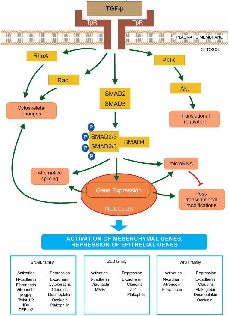
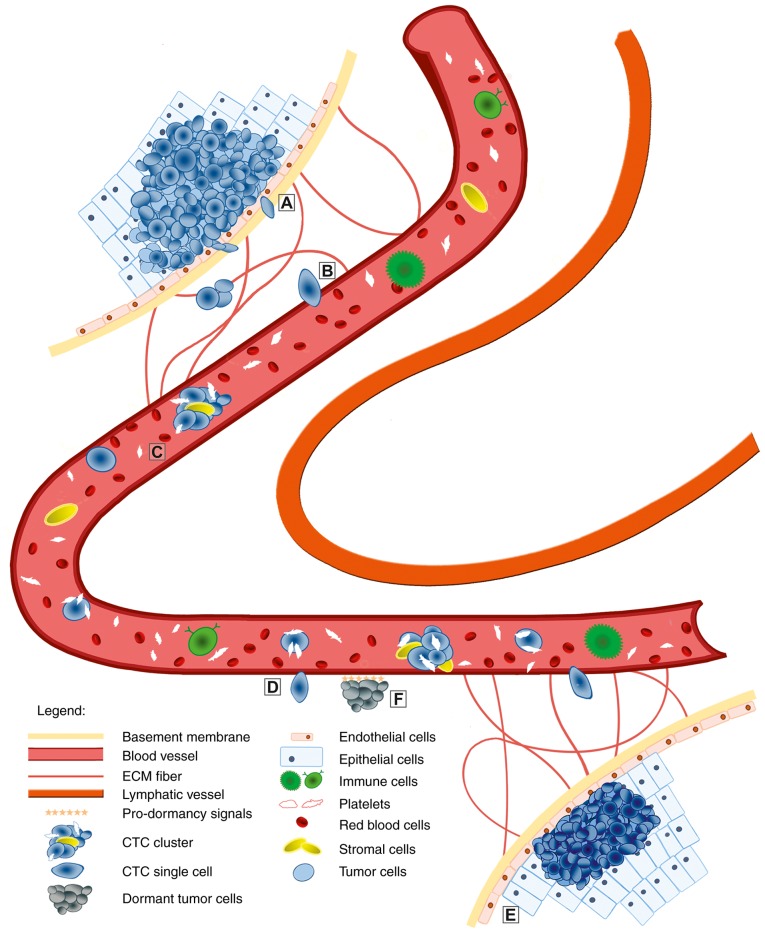
Numerous studies over the past two decades have focused on the epithelial‑to‑mesenchymal transition (EMT) and its role in the development of metastasis. Certain studies highlighted the importance of EMT in the dissemination of tumor cells and metastasis of epithelium‑derived carcinomas. Tumor metastasis is a multistep process during which tumor cells change their morphology, and start to migrate and invade distant sites. The present review discusses the current understanding of the molecular mechanisms contributing to EMT in embryogenesis, fibrosis and tumorigenesis. Additionally, the signaling pathways that initiate EMT through transcriptional factors responsible for the activation and suppression of various genes associated with cancer cell migration were investigated. Furthermore, the important role of the epigenetic modifications that regulate EMT and the reverse process, mesenchymal‑to‑epithelial transition (MET) are discussed. MicroRNAs are key regulators of various intracellular processes and current knowledge of EMT has significantly improved due to microRNA characterization. Their effect on signaling pathways and the ensuing events that occur during EMT at the molecular level is becoming increasingly recognized. The current review also highlights the role of circulating tumor cells (CTCs) and CTC clusters, and their ability to form metastases. In addition, the biological properties of different types of circulating cells based on their tumor‑forming potential are compared.


Figures

Figure 1
Figure 1
Schematic representation of EMT activation via TGF-β. The initiation and progression of EMT is regulated at all levels of macromolecule synthesis (transcription/post-transcriptional modifications and translation/post-translational modifications). TGF-β binds to its receptors (TβR) and activates EMT via the SMAD signaling pathway. SMAD2/3 are bound to the SMAD4 protein and this phosphorylated complex is translocated into the nucleus where it interacts with transcriptional factors to regulate the expression levels of EMT-specific genes. The key transcriptional factors of EMT are SNAIL, ZEB and TWIST. These are important for the activation or repression of various genes that encode proteins involved in the transition. Furthermore, TGF-β activates microRNA expression that regulates gene expression at the post-transcriptional level. By contrast, EMT transcription regulators may decrease the expression levels of specific microRNAs that affect the EMT factors important in the mesenchymal-like phenotype. Furthermore, TGF-β may induce EMT via a non-SMAD pathway, by activation of the PI3K-AKT signaling cascade, which may lead to translational regulation of EMT factors. TGF-β also initiates a decrease in the number of cell junctions and activates the cytoskeletal reorganization via RHO-GTPases. EMT, epithelial-to-mesenchymal transition; TGF-β, transforming growth factor-β; SNAIL, Snail family zinc finger transcriptional factors; TWIST, Twist family BHLH transcriptional factor; ZEB, zinc finger E-box binding homeobox; PI3K, phosphoinositide 3-kinase.
Figure 2
Figure 2
Overview of the EMT/MET process in tumor metastasis. The epithelial cells undergo a genetic transformation to become cancer cells. Genetic factors and the cancer cell microenvironment initiate the malignant conversion via the EMT process. Detached tumor cells may penetrate through the basement membrane and degrade extracellular matrix (A). Subsequently, they intravasate (B) the circulation (blood or lymphatic vessels). When CTCs or CTC clusters are in the circulation (C), only a small number survive various attacks. The surviving cells undergo transition, which is necessary during extravasation into the tissues (D). This seeding of migrating cancer cells may occur quickly. In the novel stromal environment, tumor cells form micrometastases with the ability to generate fully malignant secondary tumors (E). However, extravasated tumor cells may remain dormant (F) over a long time period before they activate the MET process and proliferate to form macrometastases. EMT, epithelial-to-mesenchymal transition; MET, mesenchymal-to-epithelial transition; CTCs, circulating tumor cells.

References

    1. Nakaya Y, Sheng G. Epithelial to mesenchymal transition during gastrulation: An embryological view. Dev Growth Differ. 2008;50:755–766. doi: 10.1111/j.1440-169X.2008.01070.x. - DOI - PubMed
    1. Qin Q, Xu Y, He T, Qin C, Xu J. Normal and disease-related biological functions of Twist1 and underlying molecular mechanisms. Cell Res. 2012;22:90–106. doi: 10.1038/cr.2011.144. - DOI - PMC - PubMed
    1. Piera-Velazquez S, Li Z, Jimenez SA. Role of endothelial- mesenchymal transition (EndoMT) in the pathogenesis of fibrotic disorders. Am J Pathol. 2011;179:1074–1080. doi: 10.1016/j.ajpath.2011.06.001. - DOI - PMC - PubMed
    1. Fidler IJ. The pathogenesis of cancer metastasis: The ‘seed and soil’ hypothesis revisited. Nat Rev Cancer. 2003;3:453–458. doi: 10.1038/nrc1098. - DOI - PubMed
    1. Książkiewicz M, Markiewicz A, Zaczek AJ. Epithelial-mesenchymal transition: A hallmark in metastasis formation linking circulating tumor cells and cancer stem cells. Pathobiology. 2012;79:195–208. doi: 10.1159/000337106. - DOI - PubMed