. 2018 Mar;41(3):1427-1436.
doi: 10.3892/ijmm.2017.3353. Epub 2017 Dec 29.

A murine model of dry eye induced by topical administration of erlotinib eye drops

Affiliations

A murine model of dry eye induced by topical administration of erlotinib eye drops

Qi-Chen Yang et al. Int J Mol Med. 2018 Mar.

Abstract

In the present study, the effects of erlotinib on mouse tear function and corneal epithelial tissue structure were investigated. Throughout the 3 weeks of treatment, no notable differences were observed in the body, eye or lacrimal gland weights of the control and experimental mice. However, in the experimental group, the tear volume and break‑up times of tear film were significantly lower following treatment with erlotinib compared with the control group. Corneal fluorescein staining in the experimental group revealed patchy staining, and the Lissamine green staining and inflammatory index were significantly higher in the experimental group at 3 weeks than in the control group. In the experimental group, the number of corneal epithelium layers increased significantly following treatment with erlotinib for 3 weeks and a significant increase in the number of vacuoles was observed compared with the control group. Treatment with erlotinib significantly increased the corneal epithelial cell apoptosis, and led to a significantly increased number of epithelial cell layers and increased keratin 10 expression. It also significantly reduced the number of conjunctival goblet cells. Transmission electron microscopy and scanning electron microscopy revealed that the corneal epithelial surface was irregular and there was a substantial reduction and partial loss of the microvilli in the experimental group. Mice treated with erlotinib also exhibited an increased protein expression of tumor necrosis factor‑α and decreased protein expression of phosphorylated‑epidermal growth factor receptor in the corneal epithelial cells. The topical application of erlotinib eye drops was revealed to induce dry eyes in mice. This is a novel method of developing a model of dry eyes in mice.


Figures

Figure 1
Figure 1
Experimental design. (A) A total of 60 mice were randomized into two groups (n=30/group). Group 1 were administered PBS eye drops, 4 times/day, whereas group 2 were administered 20 μM erlotinib 4 times/day. The (B) body, (C) eyeball and (D) extraorbital lacrimal gland weights of the mice were measured at the indicated time points. Data are presented as the mean ± standard deviation. BW, body weight.
Figure 2
Figure 2
Alterations to the ocular surface and degree of inflammation following treatment with erlotinib. (A) Representative examples of FL and LG staining following 3 weeks of treatment with either erlotinib or PBS. Images were captured using a slit‑lamp microscope with a reticule calibrated for ×16 magnification. The (B) corneal fluorescein staining score and the (C) LG scores were measured at the indicated time points. The (D) tear volume and (E) BUTs of the ocular surface were also recorded at the indicated time points in each group. (F) The overall inflammatory index for each group was determined. *P<0.05 vs. the PBS group at the same time point. Data are presented as the mean ± standard deviation. BUT, break‑up time. FL, fluorescein sodium; LG, Lissamine green.
Figure 3
Figure 3
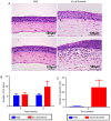
Alterations to the corneal epithelium following treatment. (A) Representative images demonstrating additional EC layers vacuole cells (black arrows) and new vessels (red arrows) in the cornea following treatment with erlotinib. (B) The number of EC layers and (C) the number of vacuole cells in the cornea epithelium was determined. Scale bar, 100 μm. Stained with hematoxylin and eosin. *P<0.05 vs. the PBS group at the same time point. EC, epithelial cell.
Figure 4
Figure 4
Representative images for PAS staining in the conjunctiva. (A) PAS staining of the forniceal conjunctiva following 3 weeks of treatment with either PBS or 20 μM erlotinib. The goblet cells (white arrows) were abundantly present in the conjunctival fornix of the PBS-treated eyes but decreased following treatment with erlotinib. (B) The average number of PAS‑positive cells in the conjunctiva was determined. The number was significantly lower in the erlotinib group compared with the PBS group following 3 weeks of treatment. Scale bar, 100 μm. *P<0.05 vs. the PBS group at the same time point. Data are presented as the mean ± standard deviation. PAS, periodic acid-Schiff.
Figure 5
Figure 5
Corneal epithelial squamous metaplasia ultrastructure. (A) Representative images from a keratin 10 (green line and red arrow) and a terminal deoxynucleotidyl transferase mediated dUTP biotin nick end labeling assay of the cornea epithelium following 3 weeks of treatment with either PBS or erlotinib. White arrows indicate apoptosis‑positive cells. (B) The level of apoptosis and keratin 10 was quantified. Few apoptotic cells were observed in the superficial layer of the corneal epithelium in the PBS group, while significantly more apoptosis was recorded in the corneal superficial and basal epithelium following treatment with erlotinib. Compared with the PBS group, there was a significant upregulation of keratin 10‑positive cells in the central cornea of the erlotinib group following 3 weeks of treatment. (C) In the PBS group, an integrated junction between epithelial cells was observed by scanning electron microscopy (upper left image, black arrow), whereas a destroyed junction (upper right image, black arrow) was observed in the erlotinib group. Following treatment with PBS for 3 weeks, the corneal epithelial microvilli were extended as digitations and arranged neatly (lower left image, blue arrow). Following treatment with erlotinib for 3 weeks, the corneal epithelial microvilli were shorter and disordered (lower right image, blue arrow). (D) The number of microvilli observed in the superficial layer of the corneal epithelium was calculated for each group. Data are presented as the mean + standard deviation. *P<0.05 vs. the PBS group at the same time point. IOD, integrated optical density.
Figure 6
Figure 6
Effect of erlotinib on TNF-α, EGFR and p-EGFR protein expression levels. (A) Following treatment for 3 weeks with either erlotinib or PBS, the protein expression levels of TNF-α were determined by western blot analysis, and the (B) density of the bands was statistically analyzed. (C) Following treatment for 3 weeks with either erlotinib or PBS, the protein expression levels of EGFR and p-EGFR were determined by western blot analysis, and the (D) density of the bands was used to statistically analyze the p-EGFR/EGFR ratio. Data are presented as mean + standard deviation. *P<0.05 vs. the PBS group at the same time point. TNF, tumor necrosis factor; EGFR, epidermal growth factor receptor; p, phosphorylated.

References

    1. Gansler T, Ganz PA, Grant M, Greene FL, Johnstone P, Mahoney M, Newman LA, Oh WK, Thomas CR, Jr, Thun MJ, et al. Sixty years of CA: A cancer journal for clinicians. CA Cancer J Clin. 2010;60:345–350. doi: 10.3322/caac.20088. - DOI - PubMed
    1. Vijayvergia N, Mehra R. Clinical challenges in targeting anaplastic lymphoma kinase in advanced non-small cell lung cancer. Cancer Chemother Pharmacol. 2014;74:437–446. doi: 10.1007/s00280-014-2517-6. - DOI - PMC - PubMed
    1. Zhou C, Wu YL, Chen G, Feng J, Liu XQ, Wang C, Zhang S, Wang J, Zhou S, Ren S, et al. Erlotinib versus chemotherapy as first-line treatment for patients with advanced EGFR mutation-positive non-small-cell lung cancer (OPTIMAL, CTONG-0802): A multicentre, open-label, randomised, phase 3 study. Lancet Oncol. 2011;12:735–742. doi: 10.1016/S1470-2045(11)70184-X. - DOI - PubMed
    1. Shepherd FA, Rodrigues Pereira J, Ciuleanu T, Tan EH, Hirsh V, Thongprasert S, Campos D, Maoleekoonpiroj S, Smylie M, Martins R, et al. Erlotinib in previously treated non-small-cell lung cancer. N Engl J Med. 2005;353:123–132. doi: 10.1056/NEJMoa050753. - DOI - PubMed
    1. Verkhivker GM. Exploring sequence-structure relationships in the tyrosine kinome space: Functional classification of the binding specificity mechanisms for cancer therapeutics. Bioinformatics. 2007;23:1919–1926. doi: 10.1093/bioinformatics/btm277. - DOI - PubMed

MeSH terms