

Pin1 facilitates isoproterenol‑induced cardiac fibrosis and collagen deposition by promoting oxidative stress and activating the MEK1/2‑ERK1/2 signal transduction pathway in rats
- PMID: 29286102
- PMCID: PMC5819929
- DOI: 10.3892/ijmm.2017.3354
Pin1 facilitates isoproterenol‑induced cardiac fibrosis and collagen deposition by promoting oxidative stress and activating the MEK1/2‑ERK1/2 signal transduction pathway in rats
Abstract
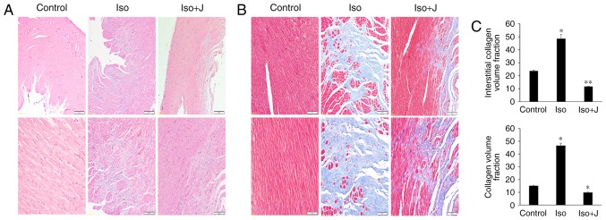
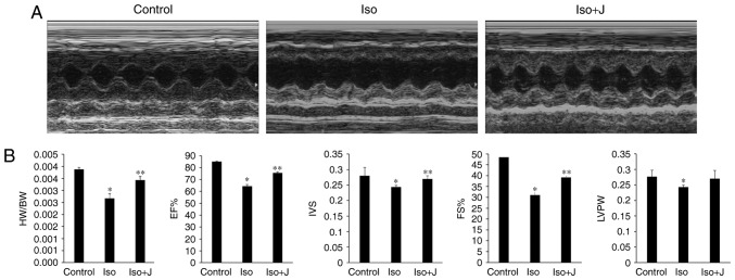
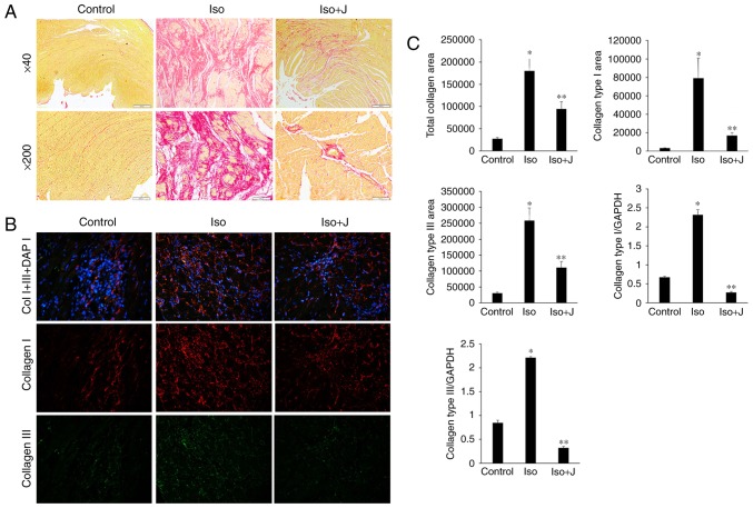
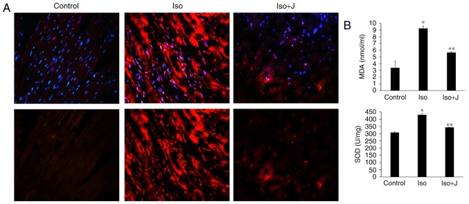
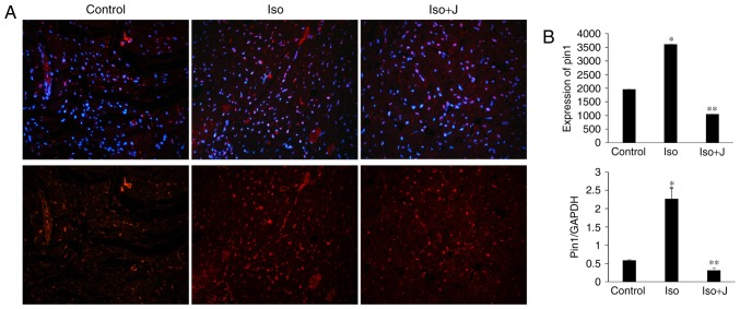
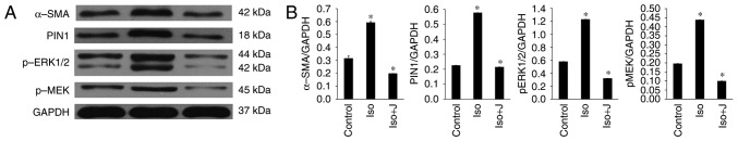
Peptidyl‑prolyl cis/trans isomerase, NIMA-interacting 1 (Pin1) is a member of a large superfamily of phosphorylation‑dependent peptidyl‑prolyl cis/trans isomerases, which not only regulates multiple targets at various stages of cellular processes, but is also involved in the pathogenesis of several diseases, including microbial infection, cancer, asthma and Alzheimer's disease. However, the role of Pin1 in cardiac fibrosis remains to be fully elucidated. The present study investigated the potential mechanism of Pin1 in isoprenaline (ISO)‑induced myocardial fibrosis in rats. The rats were randomly divided into three groups. Echocardiography was used to evaluate changes in the size, shape and function of the heart, and histological staining was performed to visualize inflammatory cell infiltration and fibrosis. Reverse transcription‑quantitative polymerase chain reaction analysis, immunohistochemistry and Picrosirius red staining were used to differentiate collagen subtypes. Additionally, cardiac‑specific phosphorylation of mitogen‑activated protein kinase kinase 1/2 (MEK1/2) and extracellular‑signal regulated protein kinase 1/2 (ERK1/2), and the activities of Pin1 and α‑smooth muscle actin (α‑SMA) and other oxidative stress parameters were estimated in the heart. The administration of ISO resulted in an increase in cardiac parameters and elevated the heart‑to‑body weight ratio. Histopathological examination of heart tissues revealed interstitial inflammatory cellular infiltrate and disorganized collagen fiber deposition. In addition, lipid peroxidation products and oxidative stress marker activity in plasma and tissues were significantly increased in the ISO‑treated rats. Western blot analysis showed significantly elevated protein levels of phosphorylated Pin1, MEK1/2, ERK1/2 and α‑SMA in remodeling hearts. Treatment with juglone following intraperitoneal injection of ISO significantly prevented inflammatory cell infiltration, improved cardiac function, and suppressed oxidative stresses and fibrotic alterations. In conclusion, the results of the present study suggested that the activation of Pin1 promoted cardiac extracellular matrix deposition and oxidative stress damage by regulating the phosphorylation of the MEK1/2‑ERK1/2 signaling pathway and the expression of α‑SMA. By contrast, the inhibition of Pin1 alleviated cardiac damage and fibrosis in the experimental models, suggesting that Pin1 contributed to the development of cardiac remodeling in ISO‑administered rats, and that the inactivation of Pin1 may be a novel therapeutic candidate for the treatment of cardiovascular disease and heart failure.
Figures
Similar articles
-
Optimized new Shengmai powder inhibits myocardial fibrosis in heart failure by regulating the rat sarcoma/rapidly accelerated fibrosarcoma/mitogen-activated protein kinase kinase/extracellular regulated protein kinases signaling pathway.J Tradit Chin Med. 2024 Jun;44(3):448-457. doi: 10.19852/j.cnki.jtcm.20240402.004. J Tradit Chin Med. 2024. PMID: 38767628 Free PMC article.
-
Tanshinone IIA attenuates heart failure via inhibiting oxidative stress in myocardial infarction rats.Mol Med Rep. 2021 Jun;23(6):404. doi: 10.3892/mmr.2021.12043. Epub 2021 Mar 31. Mol Med Rep. 2021. PMID: 33786621 Free PMC article.
-
Xinmailong mitigated epirubicin-induced cardiotoxicity via inhibiting autophagy.J Ethnopharmacol. 2016 Nov 4;192:459-470. doi: 10.1016/j.jep.2016.08.031. Epub 2016 Aug 30. J Ethnopharmacol. 2016. PMID: 27586823
-
The theatrics of collagens in the myocardium: the supreme architect of the fibrotic heart.Am J Physiol Cell Physiol. 2025 Jun 1;328(6):C1893-C1920. doi: 10.1152/ajpcell.01043.2024. Epub 2025 Apr 21. Am J Physiol Cell Physiol. 2025. PMID: 40257077 Review.
-
Juglone in Oxidative Stress and Cell Signaling.Antioxidants (Basel). 2019 Apr 5;8(4):91. doi: 10.3390/antiox8040091. Antioxidants (Basel). 2019. PMID: 30959841 Free PMC article. Review.
Cited by
-
Inhibition of MEG3 ameliorates cardiomyocyte apoptosis and autophagy by regulating the expression of miRNA-129-5p in a mouse model of heart failure.Redox Rep. 2023 Dec;28(1):2224607. doi: 10.1080/13510002.2023.2224607. Redox Rep. 2023. PMID: 37338021 Free PMC article.
-
8-Gingerol Ameliorates Myocardial Fibrosis by Attenuating Reactive Oxygen Species, Apoptosis, and Autophagy via the PI3K/Akt/mTOR Signaling Pathway.Front Pharmacol. 2021 Jul 28;12:711701. doi: 10.3389/fphar.2021.711701. eCollection 2021. Front Pharmacol. 2021. PMID: 34393792 Free PMC article.
-
Analysis of Colorectal Carcinogenesis Paradigm between Cold Constitution and Heat Constitution: Earlier ECM Collagen Deposition.Evid Based Complement Alternat Med. 2021 Jul 17;2021:5547578. doi: 10.1155/2021/5547578. eCollection 2021. Evid Based Complement Alternat Med. 2021. PMID: 34335820 Free PMC article.
-
Potential therapeutic mechanisms of Draconis Resina in cardiovascular diseases-a narrative review.Front Pharmacol. 2025 Mar 6;16:1531873. doi: 10.3389/fphar.2025.1531873. eCollection 2025. Front Pharmacol. 2025. PMID: 40115265 Free PMC article. Review.
-
Amniotic membrane mesenchymal stem cells labeled by iron oxide nanoparticles exert cardioprotective effects against isoproterenol (ISO)-induced myocardial damage by targeting inflammatory MAPK/NF-κB pathway.Drug Deliv Transl Res. 2021 Feb;11(1):242-254. doi: 10.1007/s13346-020-00788-3. Drug Deliv Transl Res. 2021. PMID: 32441012
References
-
- Barallobre-Barreiro J, Didangelos A, Schoendube FA, Drozdov I, Yin X, Fernán dez-Caggiano M, Willeit P, Puntmann VO, Aldama-López G, Shah AM, et al. Proteomics analysis of cardiac extracellular matrix remodeling in a porcine model of ischemia/reperfusion injury. Circulation. 2012;125:789–802. doi: 10.1161/CIRCULATIONAHA.111.056952. - DOI - PubMed
MeSH terms
Substances
LinkOut - more resources
Full Text Sources
Other Literature Sources
Molecular Biology Databases
Miscellaneous
