. 2018 Mar;17(3):3563-3574.
doi: 10.3892/mmr.2017.8355. Epub 2017 Dec 27.

Cigarette smoke-induced EGFR activation promotes epithelial mesenchymal migration of human retinal pigment epithelial cells through regulation of the FAK-mediated Syk/Src pathway

Affiliations

Cigarette smoke-induced EGFR activation promotes epithelial mesenchymal migration of human retinal pigment epithelial cells through regulation of the FAK-mediated Syk/Src pathway

Ga Bin Park et al. Mol Med Rep. 2018 Mar.

Abstract

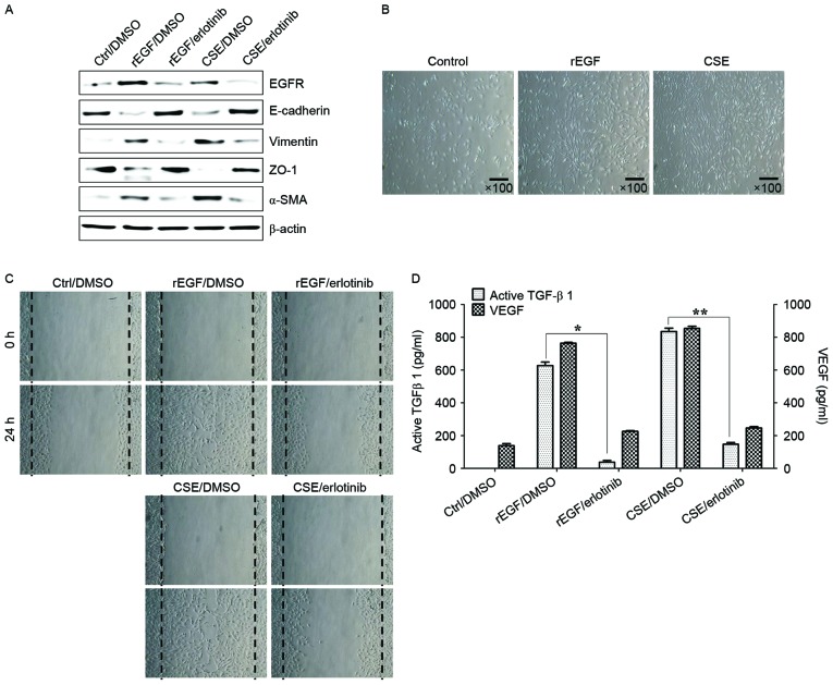
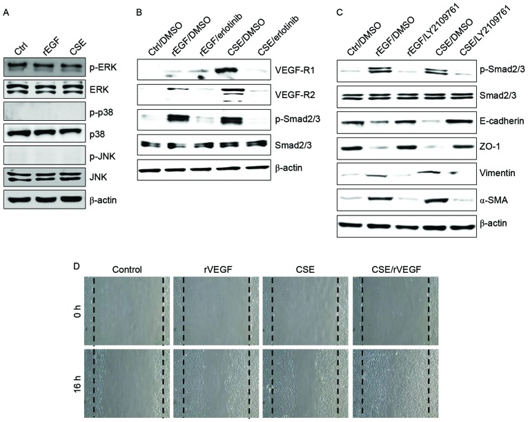
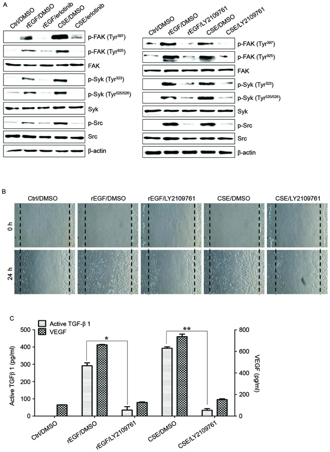
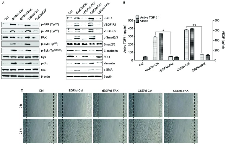
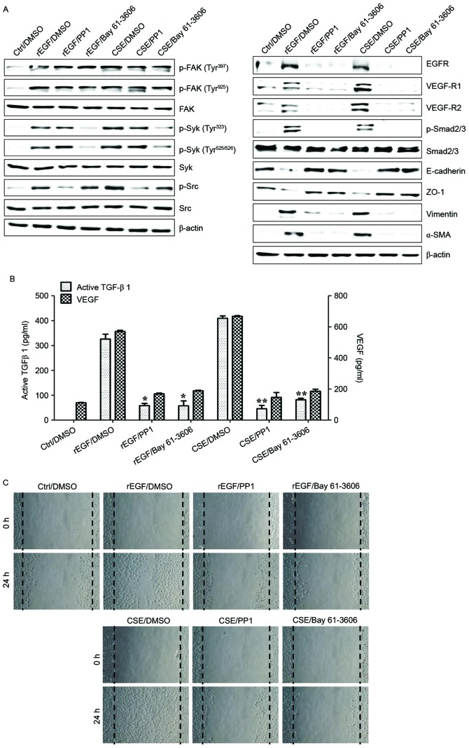
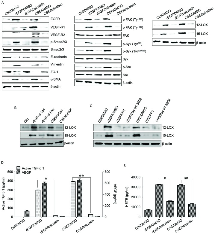
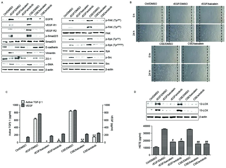
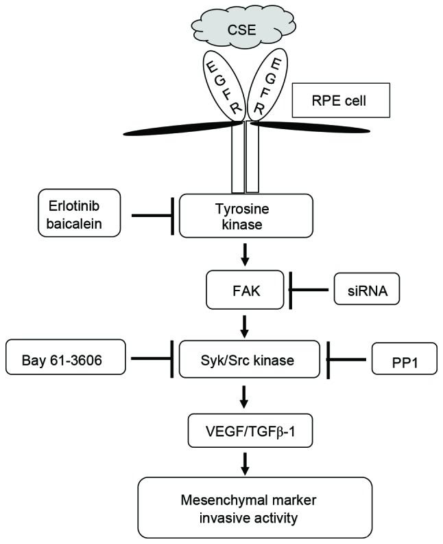
Epithelial-mesenchymal transition (EMT) of retinal pigment epithelial (RPE) cells is inevitable change of age‑related macular degeneration (AMD). Smoking is a major risk factor for the development of EMT in several diseases, including lung cancer. Cigarette smoke‑induced stress promotes the production of epidermal growth factor (EGF) in RPE cells. However, the underlying signaling pathways induced by aberrant EGF receptor (EGFR) expression in cigarette smoke-exposed RPE cells remain largely unknown. In the present study, the morphological transformation and production of EMT-associated cytokines were investigated to analyze the effect of smoking on the retina. Furthermore, EGF‑treated or cigarette smoke‑exposed RPE cells, as well as the downstream targets of EGFR, were investigated to identify the key molecules involved in EMT of cigarette smoke‑stimulated RPE cells via immunoblotting. Exposure of RPE cells to cigarette smoke extract (CSE) induced secretion of VEGF and TGF‑β1, and increased the expression of EMT markers. CSE‑mediated focal adhesion kinase (FAK) activation resulted in the phosphorylation and activation of spleen associated tyrosine kinase (Syk)/Src proto‑oncogene, non‑receptor tyrosine kinase (Src), leading to migration and invasion of RPE cells. Knockdown of FAK or pharmacological inhibition of Syk/Src abrogated CSE‑mediated VEGF and TGF‑β1 production and blocked the phosphorylation of Smad2/3 in CSE‑stimulated RPE cells. Erlotinib (an EGFR inhibitor) suppressed EGF and CSE‑mediated switch from an epithelial to mesenchymal phenotype. Baicalein, an inhi-bitor of 12/15‑lipooxygenase, also efficiently suppressed CSE‑induced EMT processes by inhibiting EGFR‑associated downstream signaling transduction. The results identified a novel signaling pathway mediated by EGFR in CSE‑activated RPE cells, and suggest baicalein as a potential new therapeutic drug for CSE‑associated retinopathy.

Keywords: retinal pigment epithelial cell; cigarette smoke; focal adhesion kinase; epidermal growth factor receptor; epithelial mesenchymal transition; 12/15-lipooxygenase.


Figures

Figure 1.
Figure 1.
CSE-mediated EGFR activation induces EMT of RPE cells through secretion of VEGF and TGF-β1. ARPE-19 cells were exposed to 5% CSE or 100 ng/ml rEGF for 4 h, subsequently washed out, and then incubated with complete medium or 50 nM erlotinib for an additional 24 h. (A) Total cell lysates were immunoblotted with the indicated antibodies. β-actin served as an internal control. (B) CSE induces mesenchymal morphology in ARPE-19 cells. Morphology was observed with an inverted phase-contrast microscope. The scale bar represents 100 µm, and photographs were taken at ×100 magnification using a digital camera. (C) Cell motility was increased by CSE as measured by a wound-healing assay. Cells were wounded (0 h) and maintained for 24 h in complete medium. Dotted lines indicate the edges of the wounds. (D) Concentrations of active TGF-β1 and VEGF in the culture supernatants were quantified by ELISA assay. Data are presented as the mean ± standard deviation of three independent experiments. Results are representative of three independent experiments. *P<0.05 and **P<0.01. Ctrl, control; DMSO, dimethyl sulfoxide; rEGF, recombinant epidermal growth factor; CSE, cigarette smoke extract; EGFR, epidermal growth factor receptor; ZO-1, tight junction protein 1; α-SMA, α-smooth muscle actin; TGF-β1, transforming growth factor-β1; VEGF, vascular endothelial growth factor.
Figure 2.
Figure 2.
CSE induces upregulation of VEGFR and activation of the transforming growth factor-β1-associated pathway. (A-C) ARPE-19 cells were exposed to 5% CSE or 100 ng/ml rEGF for 4 h, subsequently washed out, and then incubated with complete medium or 50 nM erlotinib or 100 nM LY2109761 for an additional 24 h. Total cell lysates were immunoblotted with the indicated antibodies. β-actin served as an internal control. (D) ARPE19 cells were treated with either 50 ng/ml rVEGF or 5% CSE alone, or co-treated (50 ng/ml rVEGF + 5% CSE) for 4 h, subsequently washed out, and then incubated with complete medium for an additional 24 h. Dotted lines indicate the edges of the wounds. Results are representative of three independent experiments. Ctrl, control; rEGF, recombinant epidermal growth factor; CSE, cigarette smoke extract; p, phospho; ERK, extracellular regulated kinase; JNK, c-Jun N-terminal kinase; DMSO, dimethyl sulfoxide; VEGF-R, vascular endothelial growth factor receptor; Smad, Sma- and Mad-related family; ZO-1, tight junction protein 1; α-SMA, α-smooth muscle actin; rVEGF, recombinant vascular endothelial growth factor.
Figure 3.
Figure 3.
CSE-mediated TGF-β1 activates the phosphorylation of Syk, Src and FAK for EMT in retinal pigment epithelial cells. ARPE19 cells were exposed to 5% CSE or 100 ng/ml rEGF for 4 h, subsequently washed out, and then incubated with complete medium or 50 nM erlotinib or 100 nM LY2109761 for an additional 24 h. (A) Total cell lysates of each condition were harvested and immunoblotted with the indicated antibodies. β-actin served as an internal control. (B) CSE- or rEGF-induced cell migration was decreased by LY2109761 treatment as measured by a wound healing assay. Cells were wounded (0 h) and maintained for 24 h in complete medium. Dotted lines indicate the edges of the wounds. (C) Concentrations of active TGF-β1 and VEGF in the culture supernatants were quantified by ELISA. Data are presented as the mean ± standard deviation of three independent experiments. *P<0.05 and **P<0.01. Ctrl, control; rEGF, recombinant epidermal growth factor; CSE, cigarette smoke extract; p, phospho; FAK, focal adhesion kinase; Syk, spleen associated tyrosine kinase; Src, Src proto-oncogene non-receptor tyrosine kinase; TGF-β1, transforming growth factor-β1; VEGF, vascular endothelial growth factor.
Figure 4.
Figure 4.
FAK is an upstream signal of CSE-induced Syk/Src activation and secretion of VEGF and TGF-β1. ARPE19 cells were exposed to 5% CSE or 100 ng/ml rEGF for 4 h, subsequently washed out, and then incubated with complete medium for an additional 4 h. Cell were transfected with either si-FAK-siRNA (200 nM) or si-Ctrl for 36 h prior to experiments. (A) Following transfection, protein levels of cells were detected by western blotting with the indicated antibodies. β-actin was used as a loading control. (B) Concentrations of active TGF-β1 and VEGF in the culture supernatants were quantified by ELISA assay. Data are presented as the mean ± standard deviation of three independent experiments. (C) CSE- or rEGF-induced cell migration inhibited by FAK knockdown as measured by a wound healing assay. Cells were wounded (0 h) and maintained for 24 h in complete medium. Dotted lines indicate the edges of the wounds. Results are representative of three independent experiments. *P<0.05 and **P<0.01. Ctrl, control; rEGF, recombinant epidermal growth factor; si, small interfering RNA; FAK, focal adhesion kinase; CSE, cigarette smoke extract; p, phospho; Syk, spleen associated tyrosine kinase; Src, Src proto-oncogene non-receptor tyrosine kinase; EGFR, epidermal growth factor receptor; VEGF-R, vascular endothelial growth factor receptor; Smad, Sma- and Mad-related family; ZO-1, tight junction protein 1; α-SMA, α-smooth muscle actin; TGF-β1, transforming growth factor-β1; VEGF, vascular endothelial growth factor.
Figure 5.
Figure 5.
Syk/Src activation regulates the secretion of VEGF and TGF-β1 in CSE-stimulated ARPE19 cells. ARPE19 cells were exposed to 5% CSE or 100 ng/ml rEGF for 4 h, subsequently washed out, and then incubated with complete medium, 200 nM PP1 (Src inhibitor) or 200 nM Bay 61–3606 (Syk inhibitor) for an additional 24 h. (A) Protein levels were detected by western blotting with the indicated antibodies. β-actin was used as a loading control. (B) Concentration of active TGF-β1 and VEGF in the culture supernatants were quantified by ELISA assay. Data are presented as the mean of three independent experiments, and error bars represent the standard deviation of the means. (C) PP1 or Bay 61–3606 treatment blocked CSE- or rEGF-induced cell migration as measured by wound healing assay. Cells were wounded (0 h) and maintained for 24 h in complete medium. Dotted lines indicate the edges of the wounds. Results are representative of three independent experiments. *P<0.05 and **P<0.01. Ctrl, control; DMSO, dimethyl sulfoxide; rEGF, recombinant epidermal growth factor; CSE, cigarette smoke extract; p, phospho; FAK, focal adhesion kinase; Syk, spleen associated tyrosine kinase; Src, Src proto-oncogene non-receptor tyrosine kinase; EGFR, epidermal growth factor receptor; VEGF-R, vascular endothelial growth factor receptor; Smad, Sma- and Mad-related family; ZO-1, tight junction protein 1; α-SMA, α-smooth muscle actin; TGF-β1, transforming growth factor-β1; VEGF, vascular endothelial growth factor.
Figure 6.
Figure 6.
Baicalein treatment modulates the CSE-mediated signaling pathway for EMT processes in ARPE19 cells. (A) Cells were exposed to 5% CSE or 100 ng/ml rEGF for 4 h, subsequently washed out, and then incubated with complete medium or 20 µM baicalein for an additional 24 h. (B) Cells were exposed to 5% CSE or 100 ng/ml rEGF for 4 h, subsequently washed out, and then incubated with complete medium for an additional 4 h. Cell were transfected with either FAK-siRNA (200 nM) or control-siRNA for 36 h prior to experiments. (C) Cells were exposed to 5% CSE or 100 ng/ml rEGF for 4 h, subsequently washed out, and then incubated with complete medium, 200 nM PP1 (Src inhibitor), or 200 nM Bay 61–3606, (Syk inhibitor) for an additional 24 h. Total cell lysates of each condition were harvested and immunoblotted with the indicated antibodies. β-actin served as an internal control. (D) Concentrations of active TGF-β1 and VEGF in the culture supernatants were quantified by ELISA assay. Data are presented as the mean of three independent experiments, and error bars represent the standard deviation. (E) Concentrations of HETE in the culture supernatants were quantified by ELISA assay. Data are presented as the mean of three independent experiments, and error bars represent the standard deviation. *P<0.05 and **P<0.01; #P<0.005 and ##P<0.001. Results are representative of three independent experiments. Ctrl, control; DMSO, dimethyl sulfoxide; rEGF, recombinant epidermal growth factor; CSE, cigarette smoke extract; EGFR, epidermal growth factor receptor; VEGF-R, vascular endothelial growth factor receptor; Smad, Sma- and Mad-related family; ZO-1, tight junction protein 1; α-SMA, α-smooth muscle actin; p, phospho; FAK, focal adhesion kinase; Syk, spleen associated tyrosine kinase; Src, Src proto-oncogene non-receptor tyrosine kinase; LOX, lipoxygenase; TGF-β1, transforming growth factor-β1; VEGF, vascular endothelial growth factor; HETE, hydroxyeicosatetraenoic acid.
Figure 7.
Figure 7.
Baicalein treatment modulates the CSE-mediated signaling pathway for EMT processes in HRPEpi. HRPEpi were exposed to 5% CSE or 100 ng/ml rEGF for 4 h, subsequently washed out, and then incubated with complete medium, 20 µM baicalein, or 50 nM erlotinib for an additional 24 h. (A) Total cell lysates of each condition were harvested and immunoblotted with the indicated antibodies. β-actin served as an internal control. (B) Baicalein treatment blocked CSE- or rEGF-induced cell migration as measured by wound healing assay. Cells were wounded (0 h) and maintained for 24 h in complete medium. Dotted lines indicate the edges of the wounds. Wound closure (measured after 24 h) was faster in cells treated with CSE or rEGF than in those treated with baicalein. (C) Concentrations of active TGF-β1 and VEGF in the culture supernatants were quantified by ELISA assay. Data are presented as the mean of three independent experiments, and error bars represent the standard deviation. (D) Upregulation of 12/15-LOX and production of HETE by CSE- or rEGF treatment was decreased by baicalein or erlotinib treatment. Total proteins were extracted from cell lysates and western blotting was performed for 12/15-LOX protein. Concentrations of HETE in the culture supernatants of ARPE19 cells were quantified by ELISA assay. Data are presented as the mean of three independent experiments, and error bars represent the standard deviation. Results are representative of three independent experiments. *P<0.05 and **P<0.01; #P<0.005 vs. rEGF/DMSO and ##P<0.001 vs. CSE/DMSO. HRPEpi, human primary retinal pigment epithelial cells; Ctrl, control; DMSO, dimethyl sulfoxide; rEGF, recombinant epidermal growth factor; CSE, cigarette smoke extract; EGFR, epidermal growth factor receptor; VEGF-R, vascular endothelial growth factor receptor; p, phospho; Smad, Sma- and Mad-related family; ZO-1, tight junction protein 1; α-SMA, α-smooth muscle actin; FAK, focal adhesion kinase; Syk, spleen associated tyrosine kinase; Src, Src proto-oncogene non-receptor tyrosine kinase; TGF-β1, transforming growth factor-β1; VEGF, vascular endothelial growth factor; LOX, lipoxygenase; HETE, hydroxyeicosatetraenoic acid.
Figure 8.
Figure 8.
Schematic diagram of intracellular signaling mechanisms that induce epithelial mesenchymal transition during CSE-induced aberrant EGFR activation in human RPE cells. CSE induces the expression of EGFR and increases the activity of FAK, Syk, and Src in RPE cells. These signaling molecules promote the production of TGF-β1 and VEGF and induce expression of EMT markers. CSE, cigarette smoke extract; EGFR, epidermal growth factor receptor; RPE cell, retinal pigment epithelial cell; FAK, focal adhesion kinase; siRNA, small interfering RNA; Syk, spleen associated tyrosine kinase; Src, Src proto-oncogene non-receptor tyrosine kinase; VEGF, vascular endothelial growth factor; TGF-β1, transforming growth factor-β.

Similar articles

Cited by

References

    1. Ryan SJ. Traction retinal detachment. XLIX Edward Jackson Memorial Lecture. Am J Ophthalmol. 1993;115:1–20. doi: 10.1016/S0002-9394(14)73518-4. - DOI - PubMed
    1. Charteris DG, Sethi CS, Lewis GP, Fisher SK. Proliferative vitreoretinopathy-developments in adjunctive treatment and retinal pathology. Eye (Lond) 2002;16:369–374. doi: 10.1038/sj.eye.6700194. - DOI - PubMed
    1. Charteris DG. Proliferative vitreoretinopathy: Pathobiology, surgical management, and adjunctive treatment. Br J Ophthalmol. 1995;79:953–960. doi: 10.1136/bjo.79.10.953. - DOI - PMC - PubMed
    1. Lee MY, Chou CY, Tang MJ, Shen MR. Epithelial-mesenchymal transition in cervical cancer: Correlation with tumor progression, epidermal growth factor receptor overexpression and snail up-regulation. Clin Cancer Res. 2008;14:4743–4750. doi: 10.1158/1078-0432.CCR-08-0234. - DOI - PubMed
    1. Ha GH, Kim JL, Breuer EK. TACC3 is essential for EGF-mediated EMT in cervical cancer. PLoS One. 2013;8:e70353. doi: 10.1371/journal.pone.0070353. - DOI - PMC - PubMed

MeSH terms