

Characterization of different osteoclast phenotypes in the progression of bone invasion by oral squamous cell carcinoma
- PMID: 29286135
- PMCID: PMC5802026
- DOI: 10.3892/or.2017.6166
Characterization of different osteoclast phenotypes in the progression of bone invasion by oral squamous cell carcinoma
Abstract
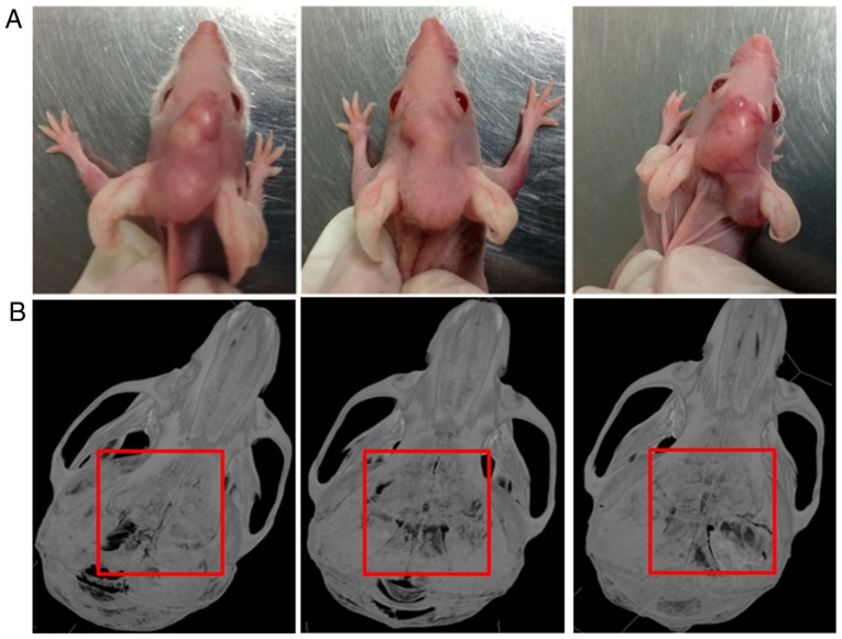
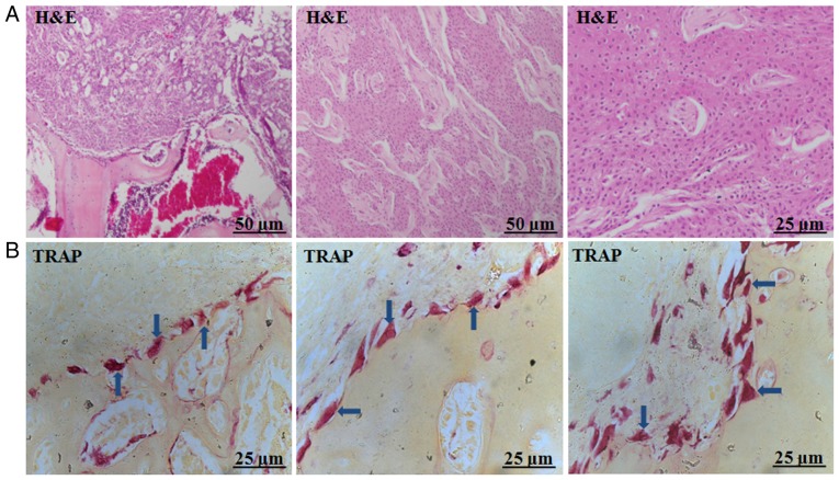
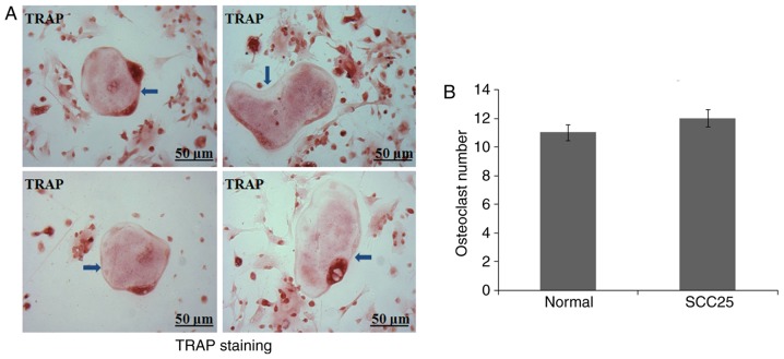
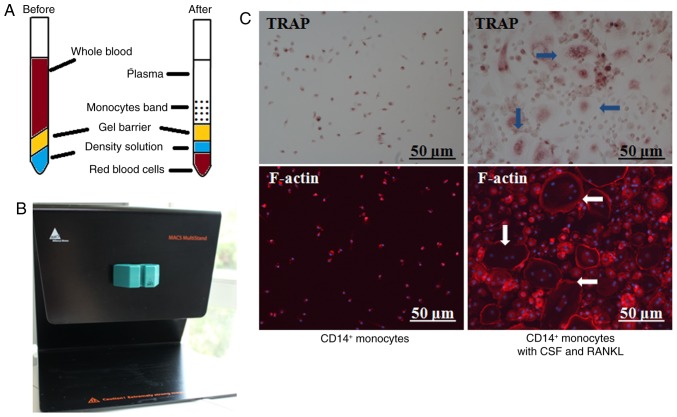
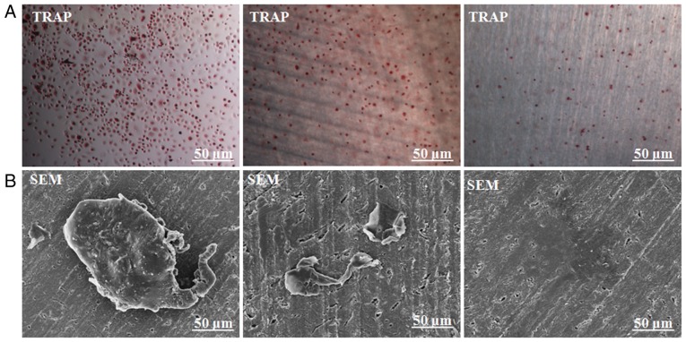
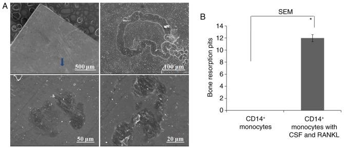
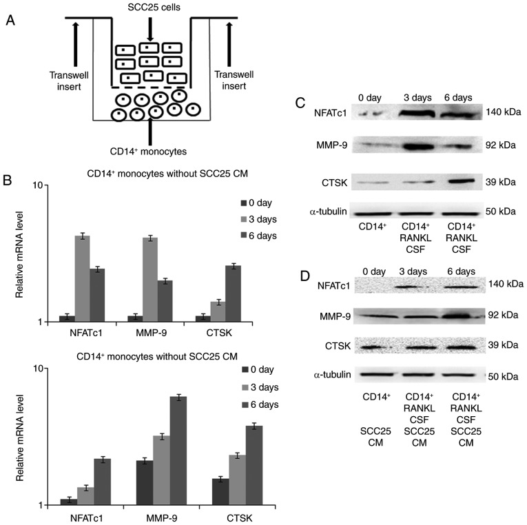
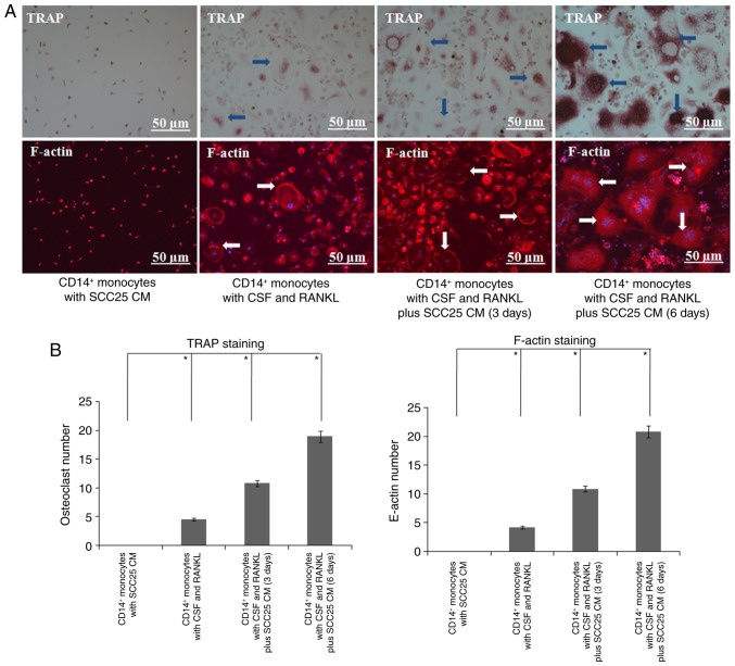
The present study aimed to characterize different phenotypes of osteoclasts in the progression of bone invasion by oral squamous cell carcinoma (OSCC). A local bone invasion model of OSCC was established by injecting SCC25 human OSCC cells into the center of calvariae in nude mice, and all mice were found to have a typical bone resorption area. Staining for tartrate-resistant acid phosphatase (TRAP) revealed various types of giant osteoclasts in the tumour-bone interface. Bone marrow cells (BMCs) were isolated from the nude mice for primary osteoclast culture, but only a few giant osteoclasts were generated. Additionally, special blood centrifuge tubes were utilized to obtain large numbers of peripheral blood mononuclear cells (PBMCs). Using magnetic activated cell sorting (MACS) and the cytokines colony-stimulating factor (CSF) and receptor activator of nuclear factor-κb ligand (RANKL), we differentiated human osteoclasts from CD14+ monocytes of PBMCs. Bone resorption was further confirmed by a bone resorption assay. Finally, Transwell inserts were used for indirect cell co-culture of SCC25 cells and CD14+ monocytes. Expression of specific osteoclast markers was detected by real-time PCR and western blotting. After co-culture for 3 and 6 days, conditioned medium (CM) of SCC25 cells stimulated the expression of osteoclast markers, and additional osteoclasts were detected through staining of TRAP and F-actin. In the present study distinct osteoclast phenotypes were observed in the established bone invasion animal model, and were confirmed using various primary osteoclast cultures. CM of OSCC cells may promote the expression of osteoclast markers and induce the differentiation of monocytes to mature osteoclasts, which can resorb adjacent bone tissue.
Figures
Similar articles
-
MCP-1 as a potential target to inhibit the bone invasion by oral squamous cell carcinoma.J Cell Biochem. 2014 Oct;115(10):1787-98. doi: 10.1002/jcb.24849. J Cell Biochem. 2014. PMID: 24905457
-
Utilization of E-cadherin by monocytes from tumour cells plays key roles in the progression of bone invasion by oral squamous cell carcinoma.Oncol Rep. 2017 Aug;38(2):850-858. doi: 10.3892/or.2017.5749. Epub 2017 Jun 23. Oncol Rep. 2017. PMID: 28656299 Free PMC article.
-
Secretion of IL-6 and IL-8 from lysophosphatidic acid-stimulated oral squamous cell carcinoma promotes osteoclastogenesis and bone resorption.Oral Oncol. 2012 Jan;48(1):40-8. doi: 10.1016/j.oraloncology.2011.08.022. Epub 2011 Sep 16. Oral Oncol. 2012. PMID: 21925926
-
Bone invasion by oral squamous cell carcinoma: Molecular alterations leading to osteoclastogenesis - a review of literature.J Craniomaxillofac Surg. 2017 Sep;45(9):1464-1471. doi: 10.1016/j.jcms.2017.04.012. Epub 2017 Apr 27. J Craniomaxillofac Surg. 2017. PMID: 28756966 Review.
-
The unique function of p130Cas in regulating the bone metabolism.Pharmacol Ther. 2022 Feb;230:107965. doi: 10.1016/j.pharmthera.2021.107965. Epub 2021 Aug 12. Pharmacol Ther. 2022. PMID: 34391790 Review.
Cited by
-
Extracellular Vesicles in Bone Homeostasis: Emerging Mediators of Osteoimmune Interactions and Promising Therapeutic Targets.Int J Biol Sci. 2022 Jun 21;18(10):4088-4100. doi: 10.7150/ijbs.69816. eCollection 2022. Int J Biol Sci. 2022. PMID: 35844790 Free PMC article. Review.
-
Hibernating bear serum hinders osteoclastogenesis in-vitro.PLoS One. 2020 Aug 27;15(8):e0238132. doi: 10.1371/journal.pone.0238132. eCollection 2020. PLoS One. 2020. PMID: 32853221 Free PMC article.
-
Unraveling non-coding RNAs in breast cancer: mechanistic insights and therapeutic potential.Med Oncol. 2024 Dec 27;42(1):37. doi: 10.1007/s12032-024-02589-x. Med Oncol. 2024. PMID: 39730979 Review.
-
Osteoclast Fusion: Physiological Regulation of Multinucleation through Heterogeneity-Potential Implications for Drug Sensitivity.Int J Mol Sci. 2020 Oct 19;21(20):7717. doi: 10.3390/ijms21207717. Int J Mol Sci. 2020. PMID: 33086479 Free PMC article. Review.
-
EZH2 engages TGFβ signaling to promote breast cancer bone metastasis via integrin β1-FAK activation.Nat Commun. 2022 May 10;13(1):2543. doi: 10.1038/s41467-022-30105-0. Nat Commun. 2022. PMID: 35538070 Free PMC article.
References
-
- Strålberg F, Kassem A, Kasprzykowski F, Abrahamson M, Grubb A, Lindholm C, Lerner UH. Inhibition of lipopolysaccharide-induced osteoclast formation and bone resorption in vitro and in vivo by cysteine proteinase inhibitors. J Leukoc Biol. 2017;101:1233–1243. doi: 10.1189/jlb.3A1016-433R. - DOI - PubMed
MeSH terms
LinkOut - more resources
Full Text Sources
Other Literature Sources
Medical
Research Materials
Miscellaneous
