Review
. 2018 Mar;41(3):1201-1212.
doi: 10.3892/ijmm.2017.3352. Epub 2017 Dec 29.

Mutual regulation of the Hippo/Wnt/LPA/TGF‑β signaling pathways and their roles in glaucoma (Review)

Affiliations
Review

Mutual regulation of the Hippo/Wnt/LPA/TGF‑β signaling pathways and their roles in glaucoma (Review)

Xin Wang et al. Int J Mol Med. 2018 Mar.

Abstract

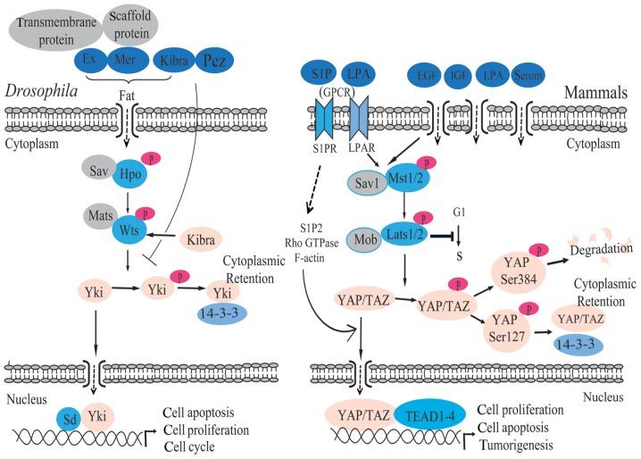
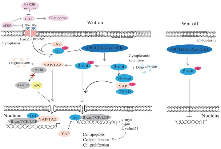
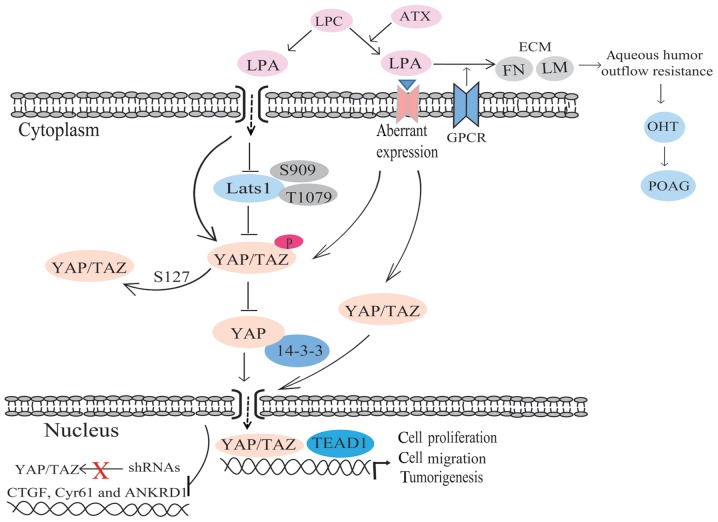
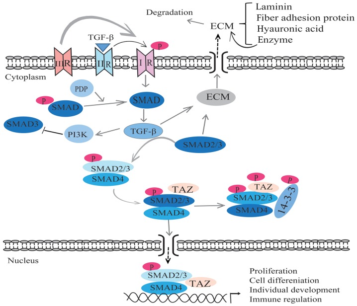
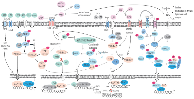
Glaucoma is the leading cause of irreversible blindness worldwide and there is no effective treatment thus far. The trabecular meshwork has been identified as the major pathological area involved. Certain signaling pathways in the trabecular meshwork, including the Wnt, lysophosphatidic acid and transforming growth factor‑β pathways, have been identified as novel therapeutic targets in glaucoma treatment. Meanwhile, it has been reported that key proteins in these pathways, particularly the primary transcription regulator Yes‑associated protein (YAP) and transcriptional co‑activator with PDZ‑binding motif (TAZ), exhibit interactions with the Hippo pathway. The Hippo pathway, which was first identified in Drosophila, has drawn great focus with regard to various aspects of studies in recent years. One role of the Hippo pathway in the regulation of organ size was indicated by more recent evidence. Defining the relevant physiological function of the Hippo pathway has proven to be extremely complicated. Studies have ascribed a role for the Hippo pathway in an overwhelming number of processes, including cell proliferation, cell death and cell differentiation. Therefore, the present review aimed to unravel the roles of YAP and TAZ in the Hippo pathway and the pathogenesis of glaucoma. Furthermore, a new and creative study for the treatment of glaucoma is provided.


Figures

Figure 1
Figure 1
Core components of the Hippo pathway. Willin is the upstream signaling molecule. Mst1/2, Lats1/2, Sav1 and Mob are the core components of the Hippo pathway. Willin stimulates Mst 1/2, then Mst 1/2 phosphorylates Lats1/2 via interaction with Sav1. Lats1/2 phosphorylates YAP/TAZ via interaction with Mob, leading to cytoplasmic retention and degradation of YAP/TAZ through phosphorylation of YAP Ser384 and binding of YAP Ser127 to 14-3-3 protein. When the kinase cascade is inactivated, phosphorylated YAP/TAZ translocate into the nucleus and generate gene expression via interaction with TEAD. YAP, Yes-associated protein; TAZ, transcriptional co-activator with PDZ-binding motif; Ex, expanded; Mer, Merlin; Sav, Salvador; Hpo, Hippo; Wts, Warts; Yki, Yorkie; Sd, scalloped; S1P, sphingosine-1-phosphate; LPA, lysophosphatidic acid; GPCR, G-protein-coupled receptor; EGF, epidermal growth factor; IGF, insulin-like growth factor; S1PR, sphingosine-1-phosphate receptor; LPAR, Lysophosphatidic acid receptor; Mst1/2, Hpo orthologs; Lats1/2, Wts orthologs; Sav1, Sav orthologs; Mob, Mats orthologs; G1, the gap before DNA replication; S, DNA synthetic phase; TEAD, transcriptional enhancer activator domain.
Figure 2
Figure 2
Regulation of the Wnt/β-catenin signaling pathway. In the off-state, APC, GSK3, Axin and CK1 form into the APC/GSK3/Axin/CK1 complex, which mediates the phosphorylation of β-catenin, repressing Wnt/β-catenin target gene transcription in the cytoplasm. In the on-state, Wnt binds with receptor Fzd and LRP5/6, forming into the Wnt/Fzd/LRP5/6 complex, and then phosphorylates and activates Dvl. Next, Dvl inhibits the activity of the APC/GSK3/Axin/CK1 complex, causing β-catenin to accumulate in the cytoplasm and then translocate into the nucleus to form a complex with the TCF/LEF family and induce gene expression. Phosphorylated β-catenin promotes degradation of TAZ via interaction with YAP/TAZ. sFRP1 inhibits β-catenin, which is inhibited by GSK3β and induces OHT, resulting in primary open-angle glaucoma. TEAD, transcriptional enhancer factor TEF-1; APC, adenomatous polyposis coli; GSK3, glycogen synthase kinase 3; CK1, casein kinase 1; LRP5/6, low-density lipoprotein receptor-related protein 5 or 6; Dvl, Dishevelled; TAZ, transcriptional co-activator with PDZ-binding motif; YAP, Yes-associated protein; sFRP1, secreted frizzled-related protein 1; OHT, ocular hypertension; sFRP1, secreted frizzled-related protein 1; OHT, ocular hypertensive; FzdR, frizzled receptors; LRP5/6R, low-density lipoprotein receptor-related protein 5 or 6; TAZ, transcriptional co-activator with PDZ-binding motif; YAP, Yes-associated protein; Dvl, dishevelled; Axin, axis inhibition protein; APC, adenomatous polyposis coli; GSK3, glycogen synthase kinase 3; CK1, casein kinase 1; β-cat, β-catenin; TCF, T-cell factor family; LEF, lymphocyte enhancer-binding factor.
Figure 3
Figure 3
Regulation of LPA in glaucoma and the mutual regulation with YAP/TAZ. LPA inhibits the activity of Lats1/2 and decreases YAP/TAZ phosphorylation, leading to the inhibition of the interaction of YAP-14-3-3 and YAP nuclear localization. LPA promotes the interaction of YAP and TEAD1. LPA promotes the accumulation of FN and LM in the human trabecular meshwork via a G-protein-coupled receptor, result in an increase in the aqueous humor outflow and OHT, finally, leading to POAG. LPA, lysophosphatidic acid; YAP, Yes-associated protein; TAZ, transcriptional co-activator with PDZ-binding motif; TEAD, transcriptional enhancer factor TEF-1; FN, fibronectin; LM, laminin; OHT, ocular hypertension; POAG, primary open-angle glaucoma; LPA, lysophosphatidic acid; ATX, autotaxin; ECM, extracellular matrix; TEAD, transcriptional enhancer activator domain; FN, fibronectin; LM, laminin; POAG, primary open-angle glaucoma.
Figure 4
Figure 4
Regulation of the TGF-β/Smad signaling pathway in glaucoma. TGF-β combines with the type II receptor, then phosphorylates the type I receptor, leading to the activation of this type I receptor. TGF-β2 promotes the synthesis of collagen protein and fiber connection protein, and inhibits the synthesis of hyaluronic acid. TGF-β2 promotes enzymes to inhibit the accumulation of the ECM. The TMC can secrete enzymes, promoting the accumulation of the ECM and increasing the aqueous outflow resistance. SMADA2/3 promotes the accumulation of the ECM in the HTM, leading to an increase in the aqueous outflow resistance and ocular hypertension, finally resulting in primary open-angle glaucoma. TGF-β, transforming growth factor-β; ECM, extracellular matrix; TAZ, transcriptional co-activator with PDZ-binding motif; TMC, trabecular meshwork cell.
Figure 5
Figure 5
Regulation amongst the Wnt/β-catenin, LPA and TGF-β/Smad signaling pathways and YAP/TAZ of the Hippo pathway. The Wnt/β-catenin, LPA and TGF-β/Smad signaling pathways are all involed in the regulation of the pathogenesis of glaucoma. LPA, lysophosphatidic acid; TGF-β, transforming growth factor-β; YAP, Yes-associated protein; TAZ, transcriptional co-activator with PDZ-binding motif; Ex, expanded; Mer, Merlin; Sav, Salvador; Hpo, Hippo; Wts, Warts; Yki, Yorkie; Sd, Scalloped; S1P, sphingosine-1-phosphate; LPA, lysophosphatidic acid; GPCR, G-protein-coupled receptor; EGF, epidermal growth factor; IGF, insulin-like growth factor; S1PR, sphingosine-1-phosphate receptor; LPAR, Lysophosphatidic acid receptor; Mst1/2, Hpo orthologs; Lats1/2, Wts orthologs; Sav1, Sav orthologs; Mob, Mats orthologs; G1, the gap before DNA replication; S, DNA synthetic phase; TEAD, transcriptional enhancer activator domain; sFRP1, Secreted frizzled-related protein 1; OHT, ocular hypertensive; FzdR, Frizzled Receptors; LRP5/6R, low-density lipoprotein receptor-related protein 5 or 6; Dvl, Dishevelled; Axin, axis inhibition protein; APC, adenomatous polyposis coli; GSK3, glycogen synthase kinase 3; CK1, casein kinase 1; β-cat, β-catenin; TCF, T-cell factor family; LEF, lymphocyte enhancer-binding factor. Axin, axis inhibition protein; ATX, autotaxin; ECM, extracellular matrix; FN, fibronectin; LM, laminin; POAG, primary open-angle glaucoma.

References

    1. Quigley HA. Open-angle glaucoma. N Engl J Med. 1993;328:1097–1106. doi: 10.1056/NEJM199304153281507. - DOI - PubMed
    1. Johnson M. ‘What controls aqueous humour outflow resistance?’. Exp Eye Res. 2006;82:545–557. doi: 10.1016/j.exer.2005.10.011. - DOI - PMC - PubMed
    1. Johnstone MA, Grant WG. Pressure-dependent changes in structures of the aqueous outflow system of human and monkey eyes. Am J Ophthalmol. 1973;75:365–383. doi: 10.1016/0002-9394(73)91145-8. - DOI - PubMed
    1. Knepper PA, Goossens W, Hvizd M, Palmberg PF. Glycosaminoglycans of the human trabecular meshwork in primary open-angle glaucoma. Invest Ophthalmol Vis Sci. 1996;37:1360–1367. - PubMed
    1. Mao W, Millar JC, Wang WH, Silverman SM, Liu Y, Wordinger RJ, Rubin JS, Pang IH, Clark AF. Existence of the canonical Wnt signaling pathway in the human trabecular meshwork. Invest Ophthalmol Vis Sci. 2012;53:7043–7051. doi: 10.1167/iovs.12-9664. - DOI - PMC - PubMed