. 2019;30(2):271-279.
doi: 10.1080/09537104.2017.1420154. Epub 2017 Dec 29.

In vivo modeling of docosahexaenoic acid and eicosapentaenoic acid-mediated inhibition of both platelet function and accumulation in arterial thrombi

Affiliations

In vivo modeling of docosahexaenoic acid and eicosapentaenoic acid-mediated inhibition of both platelet function and accumulation in arterial thrombi

Reheman Adili et al. Platelets. 2019.

Abstract

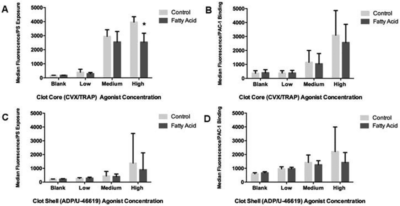
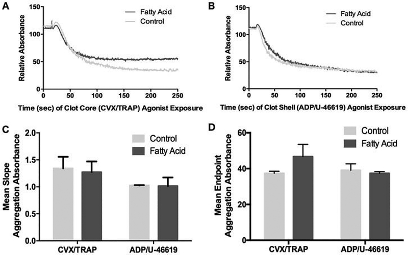
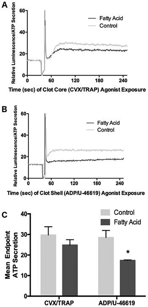
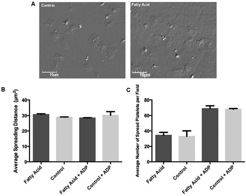
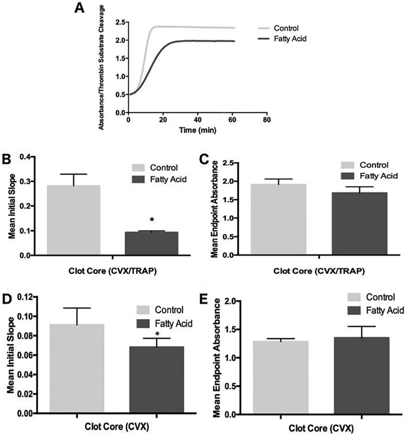
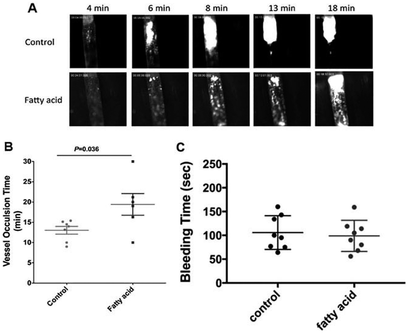
Omega-3 polyunsaturated fatty acids (n-3 PUFAs) are associated with a variety of cellular alterations that mitigate cardiovascular disease. However, pinpointing the positive therapeutic effects is challenging due to inconsistent clinical trial results and overly simplistic in vitro studies. Here we aimed to develop realistic models of n-3 PUFA effects on platelet function so that preclinical results can better align with and predict clinical outcomes. Human platelets incubated with the n-3 PUFAs docosahexaenoic acid and eicosapentaenoic acid were stimulated with agonist combinations mirroring distinct regions of a growing thrombus. Platelet responses were then monitored in a number of ex-vivo functional assays. Furthermore, intravital microscopy was used to monitor arterial thrombosis and fibrin deposition in mice fed an n-3 PUFA-enriched diet. We found that n-3 PUFA treatment had minimal effects on many basic ex-vivo measures of platelet function using agonist combinations. However, n-3 PUFA treatment delayed platelet-derived thrombin generation in both humans and mice. This impaired thrombin production paralleled a reduced platelet accumulation within thrombi formed in either small arterioles or larger arteries of mice fed an n-3 PUFA-enriched diet, without impacting P-selectin exposure. Despite an apparent lack of robust effects in many ex-vivo assays of platelet function, increased exposure to n-3 PUFAs reduces platelet-mediated thrombin generation and attenuates elements of thrombus formation. These data support the cardioprotective value of-3 PUFAs and strongly suggest that they modify elements of platelet function in vivo.

Keywords: Intravital microscopy; omega-3 fatty acids; platelet activation; platelets; thrombosis.


Figures

Figure 1.
Figure 1.
DHA- and EPA-treated platelets show mild inhibition of a-granule release and integrin activation in response to multiple agonists. Clot core agonist conditions with DHA/EPA-treated platelets were modeled using low (EC10; 0.24 pM CVX and 2.7 nM TRAP), medium (EC50; 2.4 pM CVX and 27 nM TRAP), and high (EC90; 24 pM CVX and 270 nM TRAP) concentrations of convulxin and TRAP activation conditions followed by platelet binding to a FITC-anti-P-Selectin antibody (A) or the GPIIb/IIIa active-conformation epitope antibody PAC-1 (B). Clot shell agonist conditions with DHA/EPA-treated platelets were modeled using low (2 μM ADP and 10 nM U-46619), medium (20 μM ADP and 100 nM U-46619), and high (200 μM ADP and 1.1 μM U-46619) concentrations of U46619 (a TxA2 analog) and ADP followed by binding to the P-Selectin antibody (C) or PAC-1 (D). *p < 0.01 via one-way ANOVA, n = 4, error bars represent SD. The label “Blank” refers to no agonist stimulation, showing that DHA/EPA incubation alone does not alter the platelet activation profile of these two markers.
Figure 2.
Figure 2.
DHA- and EPA-treated platelets show minimal inhibition of platelet aggregation in response to multiple agonists. Washed platelets treated with or without DHA/EPA were stimulated with EC50 doses of clot core agonists (A) or clot shell agonists (B) and measured for decreased light absorbance as a measure of platelet aggregation. A representative example is shown for each agonist condition, and (C) and (D) represent the mean of slope change and maximum aggregation, respectively, from n = 4 experiments, error bars represent SD.
Figure 3.
Figure 3.
Agonist-induced secretion of platelet dense granules is inhibited by DHA and EPA modification in response to the shell agonists, but not core agonists. Washed platelets treated with or without DHA/EPA were stimulated with EC50 doses of clot core agonists (A) or clot shell agonists (B) and measured for ATP- induced luminescence as a measure of dense granule release. A representative example is shown for (A) and (B), with (C) as a mean raw fluorescence emission from n = 4 experiments, *p = 0.001 via one-way ANOVA, error bars represent SD. The spike in (A) and (B) represents the point of agonist addition to the platelets.
Figure 4.
Figure 4.
Platelet spreading on insoluble fibrinogen is unimpaired following DHA/EPA incorporation. (A) Representative photos of platelets spread on fibrinogen, either control (left panel) or DHA/EPA-treated (right panel).(B) Quantitation of n = 50 spread platelets area using ImageJ, with or without the addition of ADP as a co- agonist. (C) Total number of platelets spread per 60× microscope field, with or without the addition of ADP as a co-agonist, n = 10 fields counted. Error bars represent SD.
Figure 5.
Figure 5.
Thrombin generation rate kinetics are impaired after DHA and EPA modification. Platelets treated with or without added DHA and EPA were stimulated with an EC50 dose of TRAP and convulxin and added to a chromogenic thrombin substrate in the presence of exogenous factor Xa and prothrombin. Increased absorbance corresponds to increased substrate cleavage by thrombin, and slope of the curves reflects thrombin generation rate kinetics. (A) A representative example of thrombin generation in human platelets treated ex vivo with DHA and EPA followed by CVX and TRAP stimulation. Averages of the mean slope (Abs/min, obtained from the steepest part of initial slop prior to leveling off) of thrombin formation (B) and maximum thrombin generation (C) for washed human platelets, n = 5. Averages of the mean slope of thrombin formation (D) and maximum thrombin generation (E) for washed murine platelets treated with 75 μg/mL CVX, n = 5. *p < 0.001, +p < 0.05 via one-way ANOVA, error bars represent SD.
Figure 6.
Figure 6.
DHA and EPA modification attenuated laser induced-thrombus formation in vivo. Thrombus formation in response to laser-induced injury on cremaster arterioles in mice fed with a control diet or DHA/EPA-rich diet. Thrombus formation at the site of vascular was monitored in real-time under intravital microscopy for 4 min. (A) Representative images of platelet (green) accumulation and fibrin formation (red) injury in mice fed with a control diet (upper panel) or DHA/EPA-rich diet (lower panel). (B) Dynamics of platelet accumulation and fibrin formation in thrombi assessed by relative mean fluorescence intensity of platelet and fibrin (P < 0.001) averaged by 10–15 thrombi per mouse three mice in each group. Data represent mean ±SEM. (C) Representative images of total platelet (green) accumulation and platelets P-selectin expression (red) within thrombi in mice fed with a control diet (upper panel) or DHA/EPA-rich diet (lower panel). (D) Dynamics of total platelet accumulation (P < 0.001) and platelet P-selectin expression (P > 0.05) in thrombi assessed by relative mean fluorescence intensity averaged by 8–10 thrombi per mouse three mice in each group. (E) Representative two-dimensional confocal images of platelet area (green) and P-selectin positive area (red) in stable thrombi recorded under confocal intravital microscopy. (F) Dynamics of total platelet accumulated area (P < 0.001) and platelet P-selectin positive area (P > 0.05) in thrombi assessed by under confocal intravital microscopy 3–4 thrombi in each mouse three mice per group. Dynamics of fluorescent intensity and surface area were analyzed Slidebook 6.0 program and compared by two-way ANOVA analysis for statistical difference.
Figure 7.
Figure 7.
DHA and EPA modification delayed the vessel occlusion in FeCI3- induced carotid artery thrombosis model. (A) Representative images of thrombosis in carotid artery in respond to FeCl3 injury. 7.5% FeCl3 was topically applied on the right carotid artery for2 min and images of thrombosis were recorded in real-time and vessel occlusion was determined by the secession of blood flow. (B) Carotid artery vessel occlusion time in control or DHA/EPA-fed mice. The mean vessel occlusion time in the control mice is 13.1 ± 0.9 min and 19.4 ± 2.7min (p = 0.036) in DHA/EPA group. Data represent mean±SEM, 6–7 mice in each group and analyzed by student’s t-test. (C) The DHA/EPA diet did not alter tail bleeding time in mice. Tail bleeding time was compared in mice with control diet and DHA/DPA diet (eight mice in each group). No significant differences were found in tail bleeding time between two groups. Data represent mean ±SEM; two-tailed unpaired t-test.

References

    1. Von Schacky C, Harris WS. Cardiovascular benefits of omega-3 fatty acids. Cardiovasc Res. 2007;73(2):310–315. - PubMed
    1. Breslow JL. n-3 fatty acids and cardiovascular disease. Am J Clin Nutr. 2006;83(6 Suppl):1477S–1482S. - PubMed
    1. Harris WS, Kennedy KF, O’Keefe JH Jr, Spertus JA. Red blood cell fatty acid levels improve GRACE score prediction of 2-yr mortality in patients with myocardial infarction. Int J Cardiol. 2013;168(1):53–59. - PMC - PubMed
    1. Farzaneh-Far R, Harris WS, Garg S, Na B, Whooley MA. Inverse association of erythrocyte n-3 fatty acid levels with inflammatory bio-markers in patients with stable coronary artery disease: the heart and soul study. Atherosclerosis. 2009;205(2):538–543. - PMC - PubMed
    1. Siscovick DS, Raghunathan TE, King I, Weinmann S, Wicklund KG, Albright J, Bovbjerg V, Arbogast P, Smith H, Kushi LH, et al. Dietary intake and cell membrane levels of long-chain n-3 polyun-saturated fatty acids and the risk of primary cardiac arrest. JAMA. 1995;274(17):1363–1367. - PubMed

MeSH terms

LinkOut - more resources