

The Tumor-Like Phenotype of Rheumatoid Synovium: Molecular Profiling and Prospects for Precision Medicine
- PMID: 29287304
- PMCID: PMC5920713
- DOI: 10.1002/art.40406
The Tumor-Like Phenotype of Rheumatoid Synovium: Molecular Profiling and Prospects for Precision Medicine
Abstract
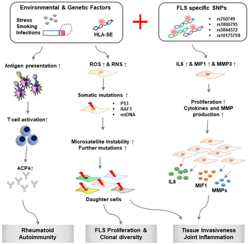
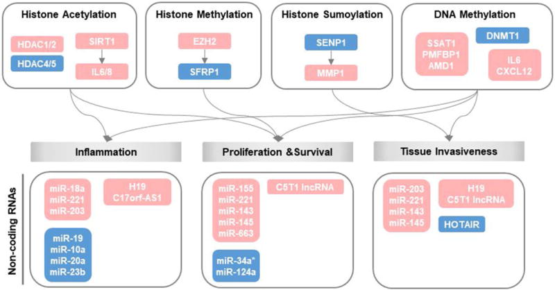
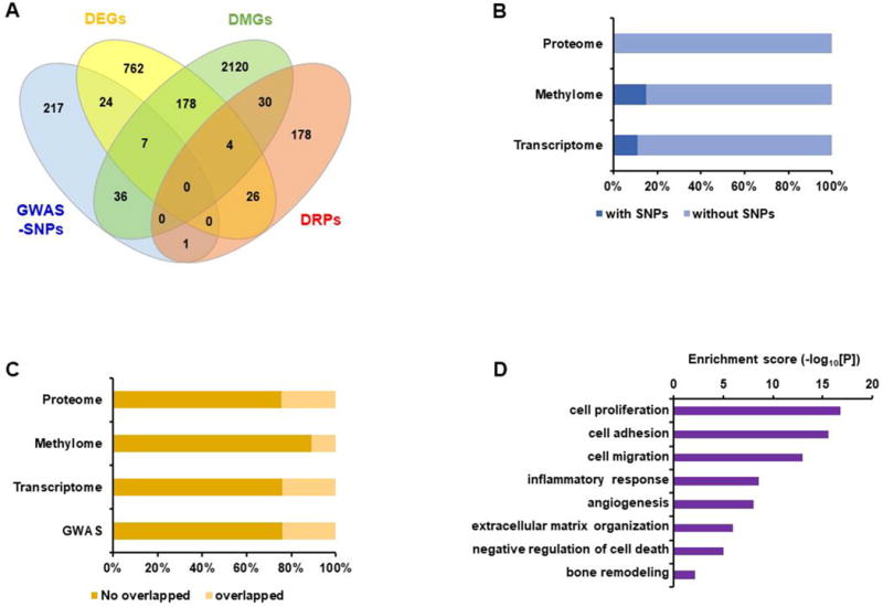
Rheumatoid arthritis (RA) is a systemic autoimmune disease characterized by destructive hyperplasia of the synovium. Fibroblast-like synoviocytes (FLS) are a major component of synovial pannus and actively participate in the pathologic progression of RA. How rheumatoid FLS acquire and sustain such a uniquely aggressive phenotype remains poorly understood. We describe the current state of knowledge of the molecular alterations in rheumatoid FLS at the genomic, epigenomic, transcriptomic, proteomic, and metabolomic levels, which offers a means to reconstruct the pathways leading to rheumatoid pannus. Such data provide new pathologic insight and suggest means to more sensitively assess disease activity and response to therapy, as well as support new avenues for therapeutic development.
© 2018, American College of Rheumatology.
Figures
Similar articles
-
GC/TOF-MS-based metabolomic profiling in cultured fibroblast-like synoviocytes from rheumatoid arthritis.Joint Bone Spine. 2016 Dec;83(6):707-713. doi: 10.1016/j.jbspin.2015.11.009. Epub 2016 Apr 25. Joint Bone Spine. 2016. PMID: 27133762
-
Rspo2 exacerbates rheumatoid arthritis by targeting aggressive phenotype of fibroblast-like synoviocytes and disrupting chondrocyte homeostasis via Wnt/β-catenin pathway.Arthritis Res Ther. 2023 Nov 9;25(1):217. doi: 10.1186/s13075-023-03198-1. Arthritis Res Ther. 2023. PMID: 37946278 Free PMC article.
-
NR1D1 modulates synovial inflammation and bone destruction in rheumatoid arthritis.Cell Death Dis. 2020 Feb 18;11(2):129. doi: 10.1038/s41419-020-2314-6. Cell Death Dis. 2020. PMID: 32071294 Free PMC article.
-
Duality of fibroblast-like synoviocytes in RA: passive responders and imprinted aggressors.Nat Rev Rheumatol. 2013 Jan;9(1):24-33. doi: 10.1038/nrrheum.2012.190. Epub 2012 Nov 13. Nat Rev Rheumatol. 2013. PMID: 23147896 Free PMC article. Review.
-
The p53 status in rheumatoid arthritis with focus on fibroblast-like synoviocytes.Immunol Res. 2021 Jun;69(3):225-238. doi: 10.1007/s12026-021-09202-7. Epub 2021 May 13. Immunol Res. 2021. PMID: 33983569 Review.
Cited by
-
Serum amyloid A expression in liver promotes synovial macrophage activation and chronic arthritis via NFAT5.J Clin Invest. 2024 Mar 1;134(5):e167835. doi: 10.1172/JCI167835. J Clin Invest. 2024. PMID: 38426494 Free PMC article.
-
Low-dose radiotherapy of osteoarthritis: from biological findings to clinical effects-challenges for future studies.Strahlenther Onkol. 2023 Dec;199(12):1164-1172. doi: 10.1007/s00066-022-02038-6. Epub 2023 Jan 5. Strahlenther Onkol. 2023. PMID: 36602569 Free PMC article. Review.
-
MiR-30e-5p deficiency exerts an inhibitory effect on inflammation in rheumatoid arthritis via regulating Atl2 expression.Arch Rheumatol. 2022 Nov 17;38(1):119-128. doi: 10.46497/ArchRheumatol.2023.9526. eCollection 2023 Mar. Arch Rheumatol. 2022. PMID: 37235116 Free PMC article.
-
Unveiling the Therapeutic Potential: Targeting Fibroblast-like Synoviocytes in Rheumatoid Arthritis.Expert Rev Mol Med. 2025 Jun 5;27:e18. doi: 10.1017/erm.2025.11. Expert Rev Mol Med. 2025. PMID: 40468839 Free PMC article. Review.
-
Non-FDG PET/CT imaging of inflammatory arthritis.Skeletal Radiol. 2025 Aug 15. doi: 10.1007/s00256-025-05006-0. Online ahead of print. Skeletal Radiol. 2025. PMID: 40815326 Review.
References
Publication types
MeSH terms
Grants and funding
LinkOut - more resources
Full Text Sources
Other Literature Sources
Medical
