. 2017 Dec 29;10(1):631.
doi: 10.1186/s13071-017-2540-7.

Molecular screening of tsetse flies and cattle reveal different Trypanosoma species including T. grayi and T. theileri in northern Cameroon

Affiliations

Molecular screening of tsetse flies and cattle reveal different Trypanosoma species including T. grayi and T. theileri in northern Cameroon

Sen Claudine Henriette Ngomtcho et al. Parasit Vectors. .

Abstract

Background: African trypanosomes are mainly transmitted through the bite of tsetse flies (Glossina spp.). The present study investigated the occurrence of pathogenic trypanosomes in tsetse flies and cattle in tsetse fly-infested areas of Northern Cameroon.

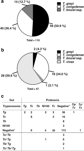
Results: Trypanosomes were identified using nested polymerase chain reaction (PCR) analysis of internal transcribed spacer 1 (ITS1) region, both by size estimation and sequencing of PCR products. Apparent density indices recorded in Gamba and Dodeo were 3.1 and 3.6 tsetse flies per trap and day, respectively. Trypanosoma prevalence infection rate for the tsetse fly gut (40%) and proboscis (19%) were recorded. Among the flies where trypanosomes were detected in the gut, 41.7% were positive for T. congolense and 14.6% for T. brucei ssp., whereas in the proboscis 36% harboured T. congolense and 62% contained T. vivax. T. grayi was highly prevalent in tsetse fly gut (58%). The most common mixed infections were the combination of T. congolense and T. grayi. Trypanosome prevalence rate in cattle blood was 6%. Among these, T. vivax represented 26%, T. congolense 35%, T. brucei ssp. 17% and T. theileri 17% of the infections. Surprisingly, in one case T. grayi was found in cattle. The mean packed cell volume (PCV) of cattle positive for trypanosomes was significantly lower (24.1 ± 5.6%; P < 0.05) than that of cattle in which trypanosomes were not detected (27.1 ± 4.9%). Interestingly, the occurrence of T. theileri or T. grayi DNA in cattle also correlated with low PCV at pathological levels.

Conclusion: This molecular epidemiological study of Trypanosoma species in Northern Cameroon revealed active foci of trypanosomes in Dodeo and Gamba. These findings are relevant in assessing the status of trypanosomosis in these regions and will serve as a guide for setting the priorities of the government in the control of the disease.

Keywords: Bodonidae; Cattle; ITS1; Northern Cameroon; Trypanosoma grayi; Trypanosoma theileri; Trypanosomosis; Tsetse fly.


Conflict of interest statement

Ethics approval

The study was conducted with the approval of the Ministère de l’élévage des pêches et des industries animales (MINEPIA) and by Mission spéciale d’éradication des glossines (MSEG) at the National and district levels, as well as the district veterinary officers in each of the study districts.

Consent for publication

Not applicable.

Competing interests

The authors declare that they have no competing interests.

Publisher’s Note

Springer Nature remains neutral with regard to jurisdictional claims in published maps and institutional affiliations.

Figures

Fig. 1
Fig. 1
Maps of the study areas. a The study area lies in the north of Cameroon, showing the administrative divisions. b Site locations: Gamba in the North Region and Dodeo, Alme and Kontcha in the Adamawa Region. Sites for flies trapping and cattle sampling are indicated. Grey shaded areas indicate the national parks Faro (near Alme) and Bénoué (near Gamba)
Fig. 2
Fig. 2
ITS1 amplicons of Trypanosoma species. a Amplicon sizes expected for amplification with generic primers ITS1-InF and ITS1-InR were calculated from available database sequences and crosschecked with sequences from screened samples. b PCR was performed with representative samples containing DNA of the indicated origin and generic (a) or specific primers (Table 1). Lane M: Marker GeneRuler 50 bp Ladder (Thermo Scientific); Lane 1: NI100 (generic primers); Lane 2: Bodonid (generic primers); Lane 3: T. vivax (generic primers); Lane 4: T. grayi (generic primers); Lane 5: T. theileri (generic primers); Lane 6: T. brucei ssp. (generic primers); Lane 7: T. grayi (specific primers); Lane 8: T. congolense (specific primers); Lane 9: T. congolense forest (specific primers); Lane 10: T. congolense forest and T. congolense kilifi (specific primers); Lane C: control without DNA template
Fig. 3
Fig. 3
The sequence of an amplicon obtained from tsetse fly gut with primers specific for T. grayi. Specific primers (TGR-In primer set) targeted against T. grayi amplified a 525 bp fragment (MG234546, Additional file 1: Table S4) from tsetse fly gut sample (ID 237-51-00211-1-40-10, G. tachinoides, Additional file 1: Table S4). The fragment was sequenced and aligned with the corresponding fragment of genomic DNA from T. grayi ANR4 (JMRU01000589)
Fig. 4
Fig. 4
Distribution of Trypanosoma species in tsetse flies. a Relative abundance of trypanosomal DNA by species in the gut. b Relative abundance of trypanosomal DNA by species in proboscis. c Correlation of trypanosomal DNA in gut and proboscis. Abbreviations: Tg, T. grayi; Tc, T. congolense; Tb, T. brucei ssp.; Tv, T. vivax. If no amplicon was detected, the fly was considered to be negative
Fig. 5
Fig. 5
Occurrence of Trypanosoma species in tsetse fly gut, proboscis and cattle blood from Dodeo. Only samples from which trypanosomal DNA was amplified were included. The percentage of each Trypanosoma species within the different tsetse fly tissues or cattle blood is displayed
Fig. 6
Fig. 6
Correlation of packed cell volume (PCV) in cattle blood with the presence of Trypanosoma DNA. a “Non-infected” vs “infected” animals. Cattle are considered “infected”, if the Trypanosoma species as a source of a PCR product was confirmed, excluding bodonid and NI100 products. All other animals were grouped as “non-infected”. The boxes indicate the corresponding 95% confidence intervals. b PCVs of individual cattle, in which DNA of the indicated parasites were detected. Dotted lines indicate the 95% confidence intervals for PCV of animals, in which no trypanosomal DNA was detected; the grey line indicates the threshold PCV of 25%. c PCVs of individual cattle, in which DNA of Bodonidae or NI100 were detected. Dotted lines indicate the 95% confidence intervals for PCV of “non-infected” animals; the grey line indicates the threshold PCV of 25%

Similar articles

Cited by

References

    1. World Health Organization . Control and surveillance of human African trypanosomiasis. Geneva: World Health Organization; 2014. - PubMed
    1. Hu C, Aksoy S. Innate immune responses regulate trypanosome parasite infection of the tsetse fly Glossina morsitans morsitans. Mol Microbiol. 2006;60:1194–1204. doi: 10.1111/j.1365-2958.2006.05180.x. - DOI - PubMed
    1. Hoare CA. Studies on Trypanosoma grayi. Trans R Soc Trop Med Hyg. 1929;23:39–56. doi: 10.1016/S0035-9203(29)90831-2. - DOI
    1. Krafsur ES. Tsetse flies: genetics, evolution, and role as vectors. Infect Genet Evol. 2009;9:124–141. doi: 10.1016/j.meegid.2008.09.010. - DOI - PMC - PubMed
    1. Baldacchino F, Muenworn V, Desquesnes M, Desoli F, Charoenviriyaphap T, Duvallet G. Transmission of pathogens by Stomoxys flies (Diptera, Muscidae): a review. Parasite. 2013;20:26. doi: 10.1051/parasite/2013026. - DOI - PMC - PubMed

Publication types

MeSH terms

LinkOut - more resources