Review
. 2018 Jul-Aug;63(4):453-479.
doi: 10.1016/j.survophthal.2017.12.008. Epub 2017 Dec 27.

Angiogenesis and lymphangiogenesis in corneal transplantation-A review

Affiliations
Review

Angiogenesis and lymphangiogenesis in corneal transplantation-A review

Wei Zhong et al. Surv Ophthalmol. 2018 Jul-Aug.

Abstract

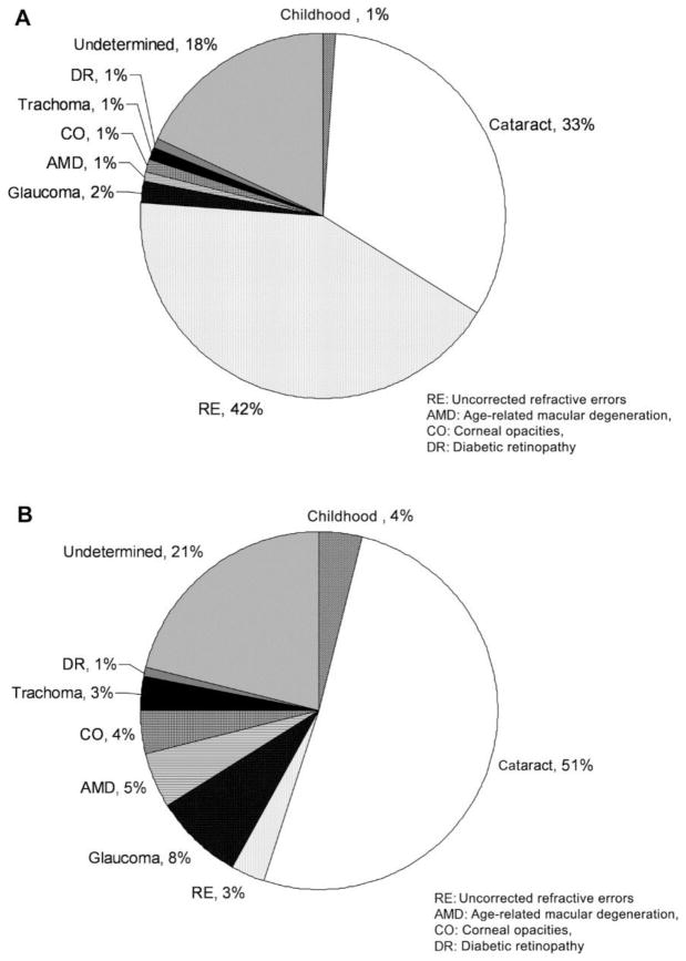
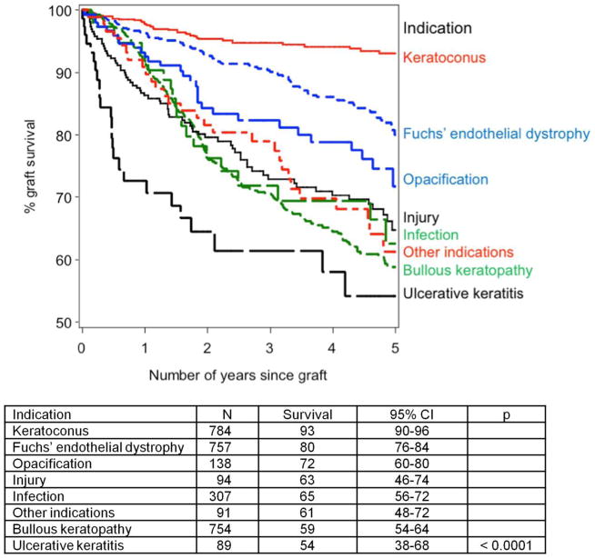
Corneal transplantation has been proven effective for returning the gift of sight to those affected by corneal disorders such as opacity, injury, and infections that are a leading cause of blindness. Immune privilege plays an important role in the success of corneal transplantation procedures; however, immune rejection reactions do occur, and they, in conjunction with a shortage of corneal donor tissue, continue to pose major challenges. Corneal immune privilege is important to the success of corneal transplantation and closely related to the avascular nature of the cornea. Corneal avascularity may be disrupted by the processes of angiogenesis and lymphangiogenesis, and for this reason, these phenomena have been a focus of research in recent years. Through this research, therapies addressing certain rejection reactions related to angiogenesis have been developed and implemented. Corneal donor tissue shortages also have been addressed by the development of new materials to replace the human donor cornea. These advancements, along with other improvements in the corneal transplantation procedure, have contributed to an improved success rate for corneal transplantation. We summarize recent developments and improvements in corneal transplantation, including the current understanding of angiogenesis mechanisms, the anti-angiogenic and anti-lymphangiogenic factors identified to date, and the new materials being used. Additionally, we discuss future directions for research in corneal transplantation.

Keywords: VEGF; VEGFR; angiogenesis; biomaterial; corneal neovascularization; corneal transplantation; lymphangiogenesis; stem cell.


Conflict of interest statement

Disclosures

No conflicts of interest, financial or otherwise, are declared by the authors.

Figures

Figure 1
Figure 1
(A) Global causes of visual impairment, inclusive of blindness, as percentage of global visual impairment in 2010. (B) Global causes of blindness as percentage of global blindness in 2010. AMD, age-related macular degeneration; CO, corneal opacities; DR, diabetic retinopathy; RE, uncorrected refractive errors. Adapted from Pascolini et al with permission from BJM)
Figure 2
Figure 2. Kaplan–Meier plot showing the effects of different indications on 5-year estimated graft survival of first corneal transplants.
(Adapted from Armitage et al with permission from IOVS)
Figure 3
Figure 3. Endothelial cell loss with aging in nondiseased eyes
Biexponential model fitted to data from cadaveric eyes (Møller-Pedersen) showing 95% prediction interval. The coefficients are shown with their respective standard error (se) and the corresponding values for t and p. The half times for the fast and slow components of the model, calculated from the relevant exponential rate constants, are 3.1 and 224 years, respectively. The residual standard deviation was 113.9 cells/mm2. Also shown for comparison are data from live subjects (Yee et al). The corresponding half times for the fast and slow components of the decay are 3.5 and 277 years, respectively. (Adapted from Armitage et al with permission from IOVS)
Figure 4
Figure 4. Endothelial cell loss after PK
Bi-exponential model fitted to data from Bourne showing 95% confidence interval (inner dotted lines) and 95% prediction interval (outer dashed lines). The coefficients are shown with their respective standard error (se) and the corresponding values for t and p. The half times for the fast and slow components of the model, calculated from the relevant exponential rate constants, are 8.6 and 257 months, respectively. The residual standard deviation was 109.6 cells/mm2. (Adapted from Armitage et al with permission from IOVS)
Figure 5
Figure 5. Schematic overview displaying
(A) a virgin cornea and (B–E) different keratoplasty procedures: (B) PK, (C) DALK, (D) DSAEK, and (E) DMEK
Figure 6
Figure 6. Average clinical finding scores for corneal rejection in all groups throughout the entire follow-up period
(a) Mean rejection scores; (b) mean opacity scores; (c) mean edema scores; and (d) mean neovascularization scores. The results obtained for the 0.5% FTY720 (Fingolimod) group, the oral FTY720 group, and the 0.05% FK506 (Tacrolimus) group were significantly lower than those obtained for the control group (all p < 0.05). Adapted from Liu et al with permission from NPG)
Figure 7
Figure 7. Fate of high-risk minor H–disparate corneal transplants in BALB/c mice (n = 14/group) that received corneal grafts from B10.D2 mice and were randomized to receive anti-CD154 or hamster Ig. Graft rejection was scored clinically
(A), and graft survival data are presented as Kaplan–Meier survival curves (B). Anti-CD154 therapy enhanced the survival of minor H–disparate grafts in high-risk transplantation (**P = 0.0001). (Adapted from Qain et al with permission from IOVS)
Figure 8
Figure 8. Fate of high-risk MHC-disparate corneal transplants in mice (n= 14/group) after anti-CD154 treatment was discontinued at week 8 after transplantation
Graft rejection was scored clinically (A), and graft survival data are presented as Kaplan–Meier survival curves (B). Only one rejection occurred after cessation of anti-CD154 therapy (**P = 0.0002). (Adapted from Qain et al with permission from IOVS)
Figure 9
Figure 9. Fate of high-risk MHC-disparate corneal transplants in BALB/c mice (n = 14/group) that received corneal grafts from BALB.b mice and were randomized to receive anti-CD154 or hamster Ig
Graft rejection was scored clinically (A), and graft survival data are presented as Kaplan–Meier survival curves (B). Anti-CD154 therapy enhanced the survival of minor H–disparate grafts in high-risk transplantation (**P = 0.0002). (Adapted from Qain et al with permission from IOVS)
Figure 10
Figure 10
Causes of corneal NV (CNV).
Figure 11
Figure 11. Rejection of corneal grafts placed into WT HSV-1–infected high-risk hosts. Wild-type BALB/c mice were infected with HSV-1 6 weeks (42 days) prior to placement of corneal grafts, which were secured with a continuous suture in this set of experiments
(a) Clinical appearance of the vascularized corneal graft bed at 6 week post HSV-1 infection; (b) corneal graft survival in vascularized corneal beds in syngraft HSK (n = 15), allograft HSK (n = 20), and allograft HSK treated with ACV (n = 10) groups compared with WT, noninfected BALB/c recipients transplanted with allograft (n = 20); (c) clinical appearance of rejected corneal allograft placed into vascularized corneal graft bed (day 3 post allograft); (d) corneal graft survival in previously infected but not vascularized (at 6 weeks post infection [p.i.]) graft beds for both allo- and syngrafts compared with WT, noninfected BALB/c recipient transplanted with allograft. (Adapted from Kuffova et al with permission from IOVS)
Figure 12
Figure 12. Generation of different transplantation models. Schematic diagram showing the generation of normal-risk (avascular), high-risk (inflamed and hemvascularized and lymphvascularized), avascular high-risk (inflamed, but avascular, recipient), and alymphatic high-risk recipient beds (inflamed and hemvascularized, but no lymphatic vessels) as transplantation models.
(Adapted from Dietrich et al with permission)
Figure 13
Figure 13
Effect of VEGF neutralization on high-risk corneal transplant survival. Animals underwent high-risk allogeneic corneal transplantation and received treatment with anti-VEGF-C, sVEGFR-3, or VEGF-trap at the time of transplantation, at 3, 7, 10, and 14 days after transplantation, and then once per week for an additional 6 weeks, or they remained untreated. The VEGF-trap treatment was most effective in increasing allograft survival (72%), though treatment with anti-VEGF-C (25%) and sVEGFR3 (11%) also significantly improved survival compared to that in the untreated control group. To create the Kaplan-Meier survival curve, graft opacity was evaluated according to an established 0 to 5+ scale by slit-lamp biomicroscopy. Scores greater than or equal to 2+ are considered rejected. Each group consisted of n=9–12 mice. **P < 0.005, *P < 0.05, error bars represent standard error of the mean (SEM). Data from one experiment of two are shown. (Adapted from Dohlman et al with permission from Wolters Kluwer)
Figure 14
Figure 14. Effect of pharmacologic neutralization of VEGF-A on survival of allogeneic cornea grafts. Panels of BALB/c mice received orthotopic transplants from C57BL/6 donors in one low-risk eye
The recipients in one panel were treated with VEGF TrapR1R2, whereas the other panel (control) received Fc-fragments only. Survival of grafts in mice treated with VEGF Trap was significantly greater than in control animals (78% vs. 40%; P < 0.05; n = 22 mice in both groups). (Adapted from Cursiefen et al with permission from IOVS)
Figure 15
Figure 15
Subconjunctival delivery of bevacizumab diminishes opacity of corneal allografts in the high-risk setting. High-risk graft beds in BALB/c mice were transplanted with C57BL/6 cornea, and mice were left untreated (n = 10) or were treated topically (n = 10) or subconjunctivally (n = 10) with bevacizumab. Corneal allografts were examined regularly to 8 weeks after transplantation by slit lamp, and graft opacity was scored using a standard grading scheme. Student's t-test was performed to evaluate statistical significance (*P < 0.05; **P < 0.01). (Adapted from Dastjerdi et al with permission from IOVS)
Figure 16
Figure 16
Subconjunctival bevacizumab promotes corneal allograft survival in the high-risk setting. High-risk graft beds in BALB/c mice were transplanted with C57BL/6 cornea, and mice were left untreated (n = 10) or were treated topically (n = 10) or subconjunctivally (n = 10) with bevacizumab. (Adapted from Dastjerdi et al with permission from IOVS)
Figure 17
Figure 17
Analysis of topical versus subconjunctival bevacizumab on the corneal NA, VC, and IA. High-risk graft beds in BALB/c mice were transplanted with C57BL/6 cornea, and mice were left untreated (n = 10) or were treated topically (n = 10) or subconjunctivally (n = 10) with bevacizumab. (A) Total area of blood vessels in each cornea was calculated and normalized to the baseline to yield the mean NA at the indicated time points to 8 weeks after transplantation. Although topical bevacizumab treatment mildly reduced NA in high-risk corneal transplantation, subconjunctival treatment resulted in a significant and marked reduction in NA at weeks 4, 6, and 8. (B) Normalized mean values for estimated blood vessel caliber at the indicated times to 8 weeks after transplantation. Although the subconjunctival treatment significantly reduced VC at week 8 (P = 0.03), topical bevacizumab appeared to have a marginal statistical difference from the control group (P = 0.05). (C) The total area of each given cornea invaded by blood vessels was calculated and normalized to yield the mean IA at the indicated times to 8 weeks after transplantation. Subconjunctival bevacizumab treatment appeared to be the only effective method to reduce IA. Student's t-test was performed to evaluate statistical significance (*P ≤ 0.05; **P < 0.01). (Adapted from Dastjerdi et al with permission from IOVS)
Figure 18
Figure 18
A, Slit-lamp image from the patient demonstrating corneal NV in the stroma crossing into the transplant at time of presentation; B, at 1 week after treatment; and C, at 1 month after treatment. (Adapted from Harooni et al with permission from Elsevier)
Figure 19
Figure 19
(A) Comparison of graft survival through 8 weeks postoperatively. The Flt23k nanoparticle group showed better survival than the PBS group (P = 0.009) until 8 weeks postoperatively and better survival than the blank nanoparticle group until 3 weeks postoperatively (P = 0.029). (B) Comparison of graft opacity grade for each week. Through 2 to 5 weeks postoperatively, the Flt23k nanoparticle group showed decreased opacification compared with the PBS group (P < 0.05). (Adapted from Cho et al with permission from IOVS) )
Figure 20
Figure 20. (A) Treatment with Flt23k nanoparticle plus triamcinolone increased graft survival compared with triamcinolone and triamcinolone plus blank nanoparticles
(P = 0.048, P = 0.020, respectively). +Sensored data, *P < 0.05, **P < 0.01. (B) The Flt23k nanoparticle plus triamcinolone group showed less total NV compared with the triamcinolone group (P = 0.000) and triamcinolone plus blank nanoparticle group (P = 0.028). The Flt23k nanoparticle plus triamcinolone group showed less graft NV compared with the triamcinolone group (P = 0.008). (C) The Flt23k nanoparticle plus triamcinolone group showed significantly less total lymphangiogenesis compared with the triamcinolone group (P = 0.043) and triamcinolone plus blank nanoparticle group (P = 0.014). The Flt23k nanoparticle plus triamcinolone group showed less graft lymphangiogenesis compared with the triamcinolone plus blank nanoparticle group (P = 0.028). (D) Representative images of NV (upper row) and lymphangiogenesis (lower row) in each group. (Adapted from Cho et al with permission from IOVS)
Figure 21
Figure 21. Survival curve for NR PK and biomicroscopic pictures from each group at 8 weeks postoperatively
VEGFR-1_MO increased graft survival compared to the PBS control treatment (P = 0.043). *P < 0.05 (Adapted from Cho et al with permission from IOVS)
Figure 22
Figure 22. Administration of exogenous endostatin promoted corneal allograft survival. (
A) Mice with corneal transplants were treated with subconjunctival injections of either endostatin or PBS (control) from postoperative day (POD) 0 to POD30. In the PBS-treated group, the allografts started to be rejected at POD27. By POD40, 6 of 10 allografts in the PBS group had been rejected. In contrast, all allografts in the endostatin-treated group survived through POD40 (p < 0.01, n = 10). (B) All syngeneic grafts survived in both treatment groups. (C) Exogenous endostatin or PBS was administered to mice from POD20, when the allografts were vascularized, to POD50. By POD60, 75% of the allografts had been rejected, and there was no significant difference between the PBS-treated group and the endostatin-treated group (p > 0.05). (Adapted from Tan et al with permission)
Figure 23
Figure 23. Lymphatic vessels in the recipient bed prior to transplantation determine graft survival
In the 2 weeks prior to transplantation (when corneal suture placement was used to induce pathologic corneal NV in the recipient bed), mice were treated with VEGF-TrapR1R2 (a [red line] and c; resulting in no blood or lymphatic vessels, but reduced inflammation in the recipient bed at the time of transplantation), the VEGFR-3 Ab mF4-31C1 (a [green line] and d; resulting in no lymphatic vessels, but only blood vessels present in the recipient bed at the time of transplantation), or the JSM6427 integrin α5β1 inhibitor (b [blue line] and e; resulting in no lymphatic vessels, but only blood vessels, present in the recipient bed at the time of transplantation). Graft survival was compared with prehemvascularized and prelymphvascularized controls (a and b [black line], f: “high-risk” recipient bed) and avascular recipient controls (a and b [dotted line], g: “low-risk” recipient bed). The graft survival was significantly better when transplants were placed into recipient beds lacking lymphatic vessels compared with beds with lymphatic vessels present at the time of transplantation (VEGF-Trap versus high-risk: p < 0.0001; VEGFR-3 versus high-risk: p < 0.0002; n = 10; JSM6427 versus high-risk: p < 0.032, n = 23; Kaplan–Meyer survival curve). (cg) Representative images of recipient corneal beds at the time of transplantation after corneas were treated with VEGF-TrapR1R2 (c), mF4-31C1 (VEGFR-3 Ab) (d), JSM6427 (e), or untreated high-risk (f) and normal-risk (g) recipient beds (original magnification ×100). Green, blood vessels; red, lymphatic vessels; arrow, prevascularized cornea. (Adapted from Dietrich at el with permission)
Figure 24
Figure 24. Overexpression of sVEGFR-3 is protective of transplant graft survival
Corneal transplant graft survival was 40.0% with subconjunctival injection of sVEGFR-3–overexpressing plasmid (pCMV.sVEGFR-3) compared with 8.3% with empty pCMV in BALB/c recipient mice (n = 9-12). **P < .05 Kaplan-Meier survival analysis.
Figure 25
Figure 25. Integrin alpha 9 blockade promoted corneal graft survival
(A) Representative images from slit-lamp examination of rejected and surviving grafts in control and treatment conditions, respectively. (B) Kaplan-Meier survival curves showing significantly higher survival rate in the treatment group. *P < 0.05. (Adapted from Kang et al with permission from IOVS)
Figure 26
Figure 26. Effect of PMab-1 on graft survival in a corneal transplantation model
(A) Fluorescence micrograph indicating mps (F4/80) and lymphangiogenesis (LYVE-1) in the PMab-1– and PBS-treated mouse corneas 7 days after corneal transplantation. (B) Quantification of the number of mps (*P = 0.0286) and of lymphangiogenesis in the corneal transplantation model assay (n = 5, each group). n.s., no significant difference. (C) Graft survival rate in mice treated with PMab-1 (n = 13) or PBS as control (n = 12). *P = 0.0259. (Adapted from Maruyama et al with permission from IOVS)
Figure 27
Figure 27. Assembly of the Boston Type I KPro device
(http://webeye.ophth.uiowa.edu/eyeforum/tutorials/Cornea-Transplant-Intro/6-kprosth.htm)

References

    1. Abdel-Naby W, Liu A, Wey N, Rosenblatt MI. Silk fibroin substrates support corneal epithelial cellular adhesion under shear stress. Invest Ophthalmol Vis Sci. 2014;55:4633–4633.
    1. Abdelfattah NS, Amgad M, Zayed AA, et al. Molecular underpinnings of corneal angiogenesis: advances over the past decade. International Journal of Ophthalmology. 2016;9:768–779. - PMC - PubMed
    1. Abdelfattah NS, Amgad M, Zayed AA, et al. Clinical correlates of common corneal neovascular diseases: a literature review. Int J Ophthalmol. 2015;8:182–193. - PMC - PubMed
    1. Abram CL, Lowell CA. The ins and outs of leukocyte integrin signaling. Annu Rev Immunol. 2009;27:339–362. - PMC - PubMed
    1. Al-Debasi T, Al-Bekairy A, Al-Katheri A, et al. Topical versus subconjunctival anti-vascular endothelial growth factor therapy (Bevacizumab, Ranibizumab and Aflibercept) for treatment of corneal neovascularization. Saudi Journal of Ophthalmology. 2017;31:99–105. - PMC - PubMed

Publication types

MeSH terms

Substances