. 2018 Mar 1:174:47-60.
doi: 10.1016/j.jprot.2017.12.016. Epub 2017 Dec 27.

Exploring the molecular complexity of Triatoma dimidiata sialome

Affiliations

Exploring the molecular complexity of Triatoma dimidiata sialome

Paula Beatriz Santiago et al. J Proteomics. .

Abstract

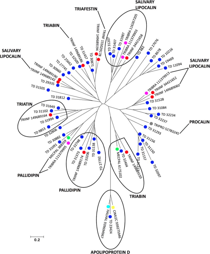
Triatoma dimidiata, a Chagas disease vector widely distributed along Central America, has great capability for domestic adaptation as the majority of specimens caught inside human dwellings or in peridomestic areas fed human blood. Exploring the salivary compounds that overcome host haemostatic and immune responses is of great scientific interest. Here, we provide a deeper insight into its salivary gland molecules. We used high-throughput RNA sequencing to examine in depth the T. dimidiata salivary gland transcriptome. From >51 million reads assembled, 92.21% are related to putative secreted proteins. Lipocalin is the most abundant gene family, confirming it is an expanded family in Triatoma genus salivary repertoire. Other putatively secreted members include phosphatases, odorant binding protein, hemolysin, proteases, protease inhibitors, antigen-5 and antimicrobial peptides. This work expands the previous set of functionally annotated sequences from T. dimidiata salivary glands available in NCBI from 388 to 3815. Additionally, we complemented the salivary analysis through proteomics (available data via ProteomeXchange with identifier PXD008510), disclosing the set complexity of 119 secreted proteins and validating the transcriptomic results. Our large-scale approach enriches the pharmacologically active molecules database and improves our knowledge about the complexity of salivary compounds from haematophagous vectors and their biological interactions.

Significance: Several haematophagous triatomine species can transmit Trypanosoma cruzi, the etiological agent of Chagas disease. Due to the reemergence of this disease, new drugs for its prevention and treatment are considered priorities. For this reason, the knowledge of vector saliva emerges as relevant biological finding, contributing to the design of different strategies for vector control and disease transmission. Here we report the transcriptomic and proteomic compositions of the salivary glands (sialome) of the reduviid bug Triatoma dimidiata, a relevant Chagas disease vector in Central America. Our results are robust and disclosed unprecedented insights into the notable diversity of its salivary glands content, revealing relevant anti-haemostatic salivary gene families. Our work expands almost ten times the previous set of functionally annotated sequences from T. dimidiata salivary glands available in NCBI. Moreover, using an integrated transcriptomic and proteomic approach, we showed a correlation pattern of transcription and translation processes for the main gene families found, an important contribution to the research of triatomine sialomes. Furthermore, data generated here reinforces the secreted proteins encountered can greatly contribute for haematophagic habit, Trypanosoma cruzi transmission and development of therapeutic agent studies.

Keywords: Chagas disease; Haematophagy; Sialome; Triatoma dimidiata; Triatominae; Vector biology.


Figures

Figure 1
Figure 1. Phylogram of the lipocalin family from Triatoma dimidiata salivary glands transcriptome
Phylogenetic tree showing the distance between members of the family is derived from the alignment of T. dimidiata CDS and other lipocalin sequences from Hemiptera identified as described in Methods section. The number at the nodes indicates percentage of bootstrap score above 70% value. The bar at the bottom represents 10% amino acid substitution. The colored circles identify the species whose sequences were used: blue, T. dimidiata; red, Triatoma infestans; green, Meccus pallidipennis; magenta, Triatoma brasiliensis; grey, Triatoma protracta; yellow, Cimex lectularius; cyan, Halyomorpha halys.
Figure 2
Figure 2. The odorant binding protein (OBP) from Triatoma dimidiata salivary glands transcriptome
ClustalW alignment of OBP members from T. dimidiata salivary transcriptome (TD_3904 and TD_4743) and other OBP sequences from Hemiptera identified as described in Methods section: Triatoma brasiliensis (gi 1026942666), T. brasiliensis (gi 1026942662), Apolygus lucorum (gi 1007629701), Adelphocoris lineolatus (gi 270000360), Euschistus heros (gi 299474028) and Lygus lineolaris (gi 573006010). The alignment indicates conserved residues in black and similar residues in grey background. The blue bar indicates the signal peptide indicative of secretion and the hashtag above indicate the position of the conserved cysteine residues.
Figure 3
Figure 3. Phylogram of the antigen-5 family from Triatoma dimidiata salivary glands transcriptome
Phylogenetic tree showing the distance between members of the family is derived from the alignment of T. dimidiata CDS and other antigen-5 sequences from Insecta identified as described in Methods section. The number at the nodes indicates percentage of bootstrap score above 70% value. The bar at the bottom represents 20% amino acid substitution.
Figure 4
Figure 4. Phylogram of the inositol polyphosphate 5-phosphatase (IPP) family from Triatoma dimidiata salivary glands transcriptome
Phylogenetic tree showing the distance between members of the family is derived from the alignment of T. dimidiata CDS and other IPPs sequences from Insecta identified as described in Methods section. The number at the nodes indicates percentage of bootstrap score above 70% value. The bar at the bottom represents 10% amino acid substitution.
Figure 5
Figure 5. Apyrase/5′nucleotidase from Triatoma dimidiata salivary glands transcriptome
(A) ClustalW alignment of apyrase/5′nucleotidase members from T. dimidiata salivary transcriptome (TD_31776 and TD_31514) and other apyrase/5′nucleotidase sequences from Insecta: T. infestans (gi 34481604), Cyphomyrmex costatus (gi 1009376035), Atta colombica (gi 1009353741), Trachymyrmex cornetzi (gi 1009393714), Trachymyrmex zeteki (gi 1012981877) and Harpegnathos saltator (gi 307202233). The alignment indicates conserved residues in black and similar residues in grey background. The blue bar indicates the signal peptide indicative of secretion, while the green indicates the 5′nucleotide superfamily located in C terminal domain. The hashtag shows the metal binding site residues and the asterisk shows the active site residues. (B) Phylogenetic tree showing the distance between members of the family is derived from the alignment of T. dimidiata CDS and other apyrase/5′nucleotidase sequences from Insecta identified as described in Methods section. The number at the nodes indicates percentage of bootstrap score above 70% value. The bar at the bottom represents 10% amino acid substitution.
Figure 6
Figure 6. Phylogram of the serine proteases from Triatoma dimidiata salivary glands transcriptome
Phylogenetic tree showing the distance between members of the family is derived from the alignment of T. dimidiata CDS and other serine proteases sequences from Insecta identified as described in Methods section. The number at the nodes indicates percentage of bootstrap score above 70% value. The bar at the bottom represents 20% amino acid substitution. The colored circles identify the species whose sequences were used: blue, T. dimidiata; red, Hemiptera order; and green, Hymenoptera order.
Figure 7
Figure 7. The serine protease from Triatoma dimidiata salivary glands transcriptome
ClustalW alignment of serine protease member from T. dimidiata salivary gland transcriptome (TD_4045) and other serine proteases sequences from Insecta: Cimex lectularius (gi 939277320), Halyomorpha halys (gi 939641366), Diuraphis noxia (gi 985385408), Copidosoma floridanum (gi 936576698) and Apis dorsata (gi 572262138). The alignment indicates conserved residues in black and similar residues in grey background. The blue bar indicates the signal peptide indicative of secretion. The triangle indicates the cleavage site, the circles indicate the active site and the diamonds indicate the substrate binding sites.
Figure 8
Figure 8. Phylogram of the kazal-type protease inhibitors from Triatoma dimidiata salivary glands transcriptome
Phylogenetic tree showing the distance between members of the family is derived from the alignment of T. dimidiata CDS and other kazal-type protease inhibitors sequences from Insecta identified as described in Methods section. The number at the nodes indicates percentage of bootstrap score above 70% value. The bar at the bottom represents 20% amino acid substitution.
Figure 9
Figure 9. Phylogram of the serpin inhibitors from Triatoma dimidiata salivary glands transcriptome
Phylogenetic tree showing the distance between members of the family is derived from the alignment of T. dimidiata CDS and other serpin inhibitors sequences from Insecta identified as described in Methods section. The number at the nodes indicates percentage of bootstrap score above 70% value. The bar at the bottom represents 10% amino acid substitution.
Figure 10
Figure 10. The pacifastin inhibitors from Triatoma dimidiata salivary glands transcriptome
Arrangement of pacifastin domain showing the residues position of the conserved inhibitory subunit, composed by C1 X9–12 C2 N X C3 X C4 X2–3 G X3–4 C5 T X3 C6, containing the six cysteines residues responsible for the intra-chain disulfide bridges: C1:C4, C2:C6, C3:C5. The T. dimidiata CDS contain pacifastin domain are TD_27731, TD_354, TD_3748 and TD_7118.
Figure 11
Figure 11. Triatoma dimidiata salivary transcriptome/proteome correlation
The diagram shows positive linear relationship between reads and proteins abundance (p<0.0001).

References

    1. Bern C. Chagas’ Disease. N Engl J Med. 2015;373(5):456–66. - PubMed
    1. Andrade DV, Gollob KJ, Dutra WO. Acute chagas disease: new global challenges for an old neglected disease. PLoS Negl Trop Dis. 2014;8(7):e3010. - PMC - PubMed
    1. Dorn PL, Monroy C, Curtis A. Triatoma dimidiata (Latreille, 1811): a review of its diversity across its geographic range and the relationship among populations. Infect Genet Evol. 2007;7(2):343–52. - PubMed
    1. Ramírez CJ, et al. Genetic structure of sylvatic, peridomestic and domestic populations of Triatoma dimidiata (Hemiptera: Reduviidae) from an endemic zone of Boyaca. Colombia Acta Trop. 2005;93(1):23–9. - PubMed
    1. Guhl F. Chagas disease in Andean countries. Mem Inst Oswaldo Cruz. 2007;102(Suppl 1):29–38. - PubMed

Publication types

LinkOut - more resources