. 2017 Dec 1;58(14):6489-6499.
doi: 10.1167/iovs.16-20673.

PI3K/Akt Pathway: A Role in δ-Opioid Receptor-Mediated RGC Neuroprotection

Affiliations

PI3K/Akt Pathway: A Role in δ-Opioid Receptor-Mediated RGC Neuroprotection

Shahid Husain et al. Invest Ophthalmol Vis Sci. .

Abstract

Purpose: This study examines the role of PI3K/Akt pathway in δ-opioid receptor agonist (SNC-121)-induced RGC neuroprotection in a chronic glaucoma rat model.

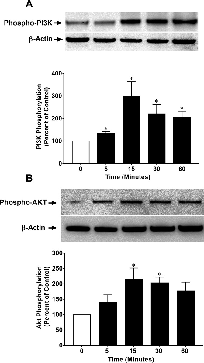
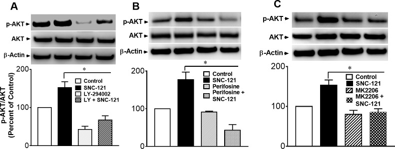
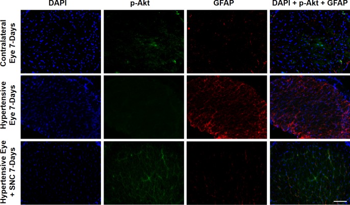
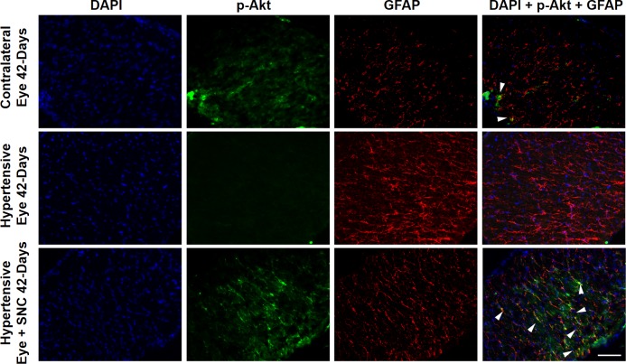
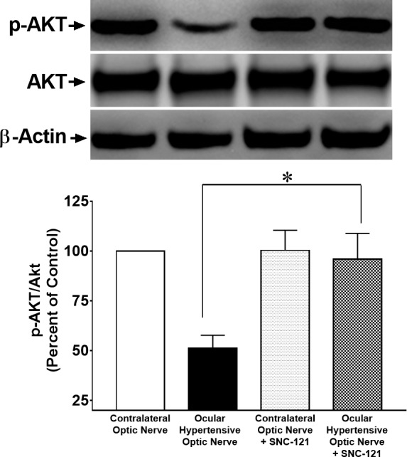
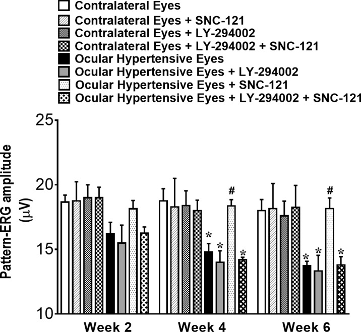
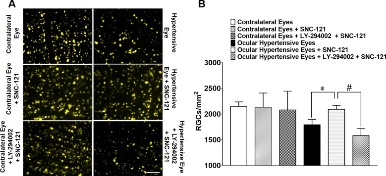
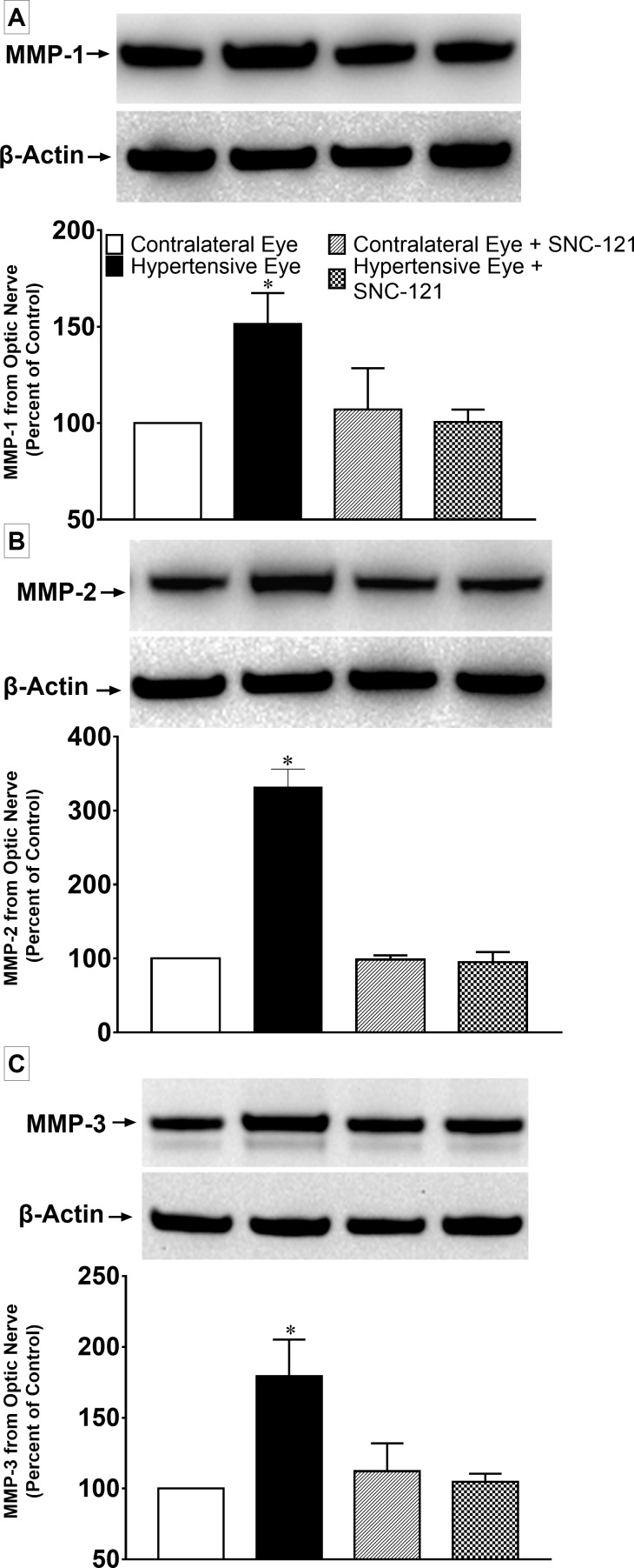
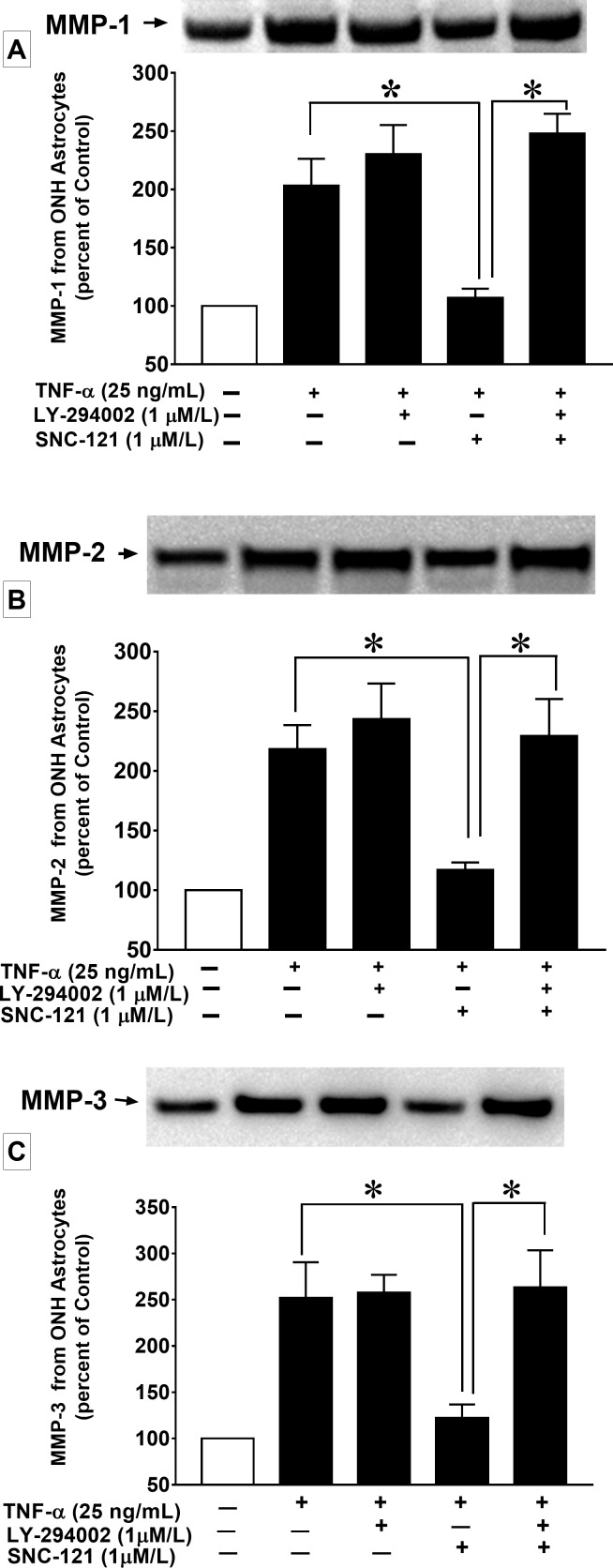
Methods: Injecting hypertonic saline into the limbal veins of Brown Norway rats elevated IOP. Rats were treated either with 1 mg/kg SNC-121 or 3 mg/kg 2-(4-morpholinyl)-8-phenyl-1(4H)-benzopyran-4-one hydrochloride (LY-294002; PI3K/Akt inhibitor) plus SNC-121 once daily for 7 days. Pattern ERGs were recorded in response to contrast reversal of patterned visual stimuli. Retinal ganglion cells (RGC) were visualized by Fluorogold retrograde labeling. Optic nerve head (ONH) astrocytes were pretreated with PI3K/Akt inhibitors for 30 minutes followed by 1-μM SNC-121 treatment. Changes in matrix metalloproteinases (MMP-1, -2, and -3) production and PI3K/Akt activation in optic nerve and TNF-α treated ONH astrocytes were measured by immunohistochemistry and Western blotting.

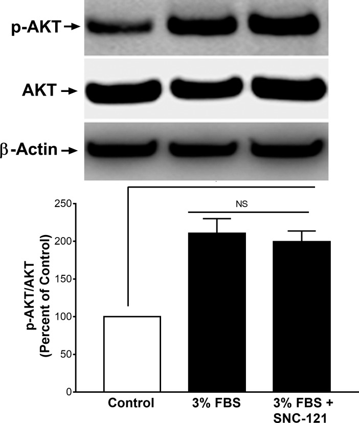
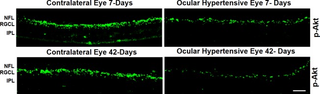
Results: SNC-121 activates the PI3K/Akt pathway in ONH astrocytes and the retina. In ONH astrocytes, SNC-121-induced Akt activation was fully inhibited by PI3K/Akt inhibitors. A sustained decline (7-42 days post injury) in Akt activation was seen in the ocular-hypertensive retina and optic nerve. This decline is reversed to normal levels by 1-mg/kg intraperitoneally (i.p.) SNC-121 treatment. Both pattern ERG amplitudes and RGC numbers were reduced in ocular hypertensive eyes, which were significantly increased in SNC-121-treated animals. Interestingly, SNC-121-induced increase in pattern-ERG amplitudes and RGC numbers were inhibited in LY-294002 pretreated animals. Additionally, SNC-121 treatment inhibited MMP-1, -2, and -3 production from the optic nerve of ocular hypertensive rats and TNF-α-treated ONH astrocytes.

Conclusions: PI3K/Akt pathway plays a crucial role in SNC-121-mediated RGC neuroprotection against glaucomatous injury.


Figures

Figure 1
Figure 1
Effects of 3% FBS and SNC-121 on Akt phosphorylation in ONH astrocytes. Cells were treated with 3% FBS alone or 3% FBS + SNC-121 (1 μM) for 30 minutes. After incubation, cells were lysed using buffer (20 mM β-glycerophosphate, pH 7.5 containing 1% Triton X-100, 20-mM EGTA, 20-mM NaF, 15-mM MgCl2, 1-mM Sodium Orthovanadate, and 0.3% Mercaptoethanol). Cell lysates (15-μg proteins) were analyzed by Western blotting using selective anti–phospho-Akt and anti-Akt antibodies. The signal was captured using enhanced chemiluminescent reagent and the Biorad Versadoc imaging system. Data expressed as mean ± SE. *P < 0.05; n = 9.
Figure 2
Figure 2
SNC-121–induced activation of PI3K (A) and Akt (B) in ONH astrocytes. ONH astrocytes were starved in serum-free medium for 16 hours. Cells were then treated with SNC-121 (1 μM) for the indicated time intervals. Cell lysates (15-μg proteins) were analyzed by Western blotting using selective anti–phospho-PI3K or anti-phospho-Akt antibodies. The band intensities were captured using enhanced chemiluminescent reagent and the Biorad Versadoc imaging system. Data are expressed as mean ± SE. *P < 0.05; n = 5–9 for phospho-PI3K and n = 7–13 for phospho-Akt.
Figure 3
Figure 3
Effects of PI3K/Akt inhibitor LY-294002 (A), Akt inhibitor, perifosine (B), and Akt inhibitor, MK-2206 (C) on SNC-121-induced phosphorylation of Akt in ONH astrocytes. ONH astrocytes were starved in serum-free medium for 16 hours followed by SNC-121 (1 μM) treatment for 30 minutes. ONH astrocytes were preincubated with LY-294002 (1 μM/L), perifosine (10 μM/L), or MK-2206 (100 nM/L) to block Akt phosphorylation for 30 minutes before SNC-121 treatment. Cells lysates (15-μg proteins) were analyzed by Western blotting using selective anti–phospho-Akt and anti-Akt antibodies. The band intensities were captured using enhanced chemiluminescent reagent and the Biorad Versadoc imaging system. Data are expressed as mean ± SE. *P < 0.05; n = 5–9, ANOVA.
Figure 4
Figure 4
Both contralateral and ocular hypertensive eyes of Brown Norway rats were removed on day 7 and 42, post injury. Contralateral eyes were used as the control. Cryosections were immunostained by anti–phospho-Akt antibodies. We did not see any positive staining when primary antibodies were omitted (not shown). Data are a representation of at least four independent experiments. Comparable staining for phospho-Akt was seen in at least eight animals. NFL, nerve fiber layer; RGCL, retinal ganglion cells layer; IPL, inner plexiform layer.
Figure 5
Figure 5
Changes in Akt phosphorylation in the optic nerve of contralateral, ocular hypertensive, and SNC-121–treated ocular hypertensive eyes. Optic nerves of untreated and SNC-121–treated animals were collected at day 7, post injury. Coronal sections of optic nerve were analyzed for phospho-Akt by immunohistochemistry using anti–phospho-Akt and anti-GFAP antibodies. Data are a representation of at least four independent experiments. Scale bar: 20 μm.
Figure 6
Figure 6
Co-localization of p-Akt and GFAP and changes in Akt phosphorylation and GFAP expression in the optic nerve of contralateral, ocular hypertensive, and SNC-121–treated ocular-hypertensive eyes. Optic nerves of untreated and SNC-121–treated animals were collected at day 42, post injury and analyzed for phospho-Akt and GFAP by immunohistochemistry using anti–phospho-Akt (1:100) and anti-GFAP (1:200) antibodies. Co-localization of phosphor-Akt and GFAP is indicated by arrowhead. Data are a representation of at least four independent experiments. Scale bar: 20 μm.
Figure 7
Figure 7
Changes in Akt phosphorylation in the optic nerve of ocular-hypertensive rats in response to the injury and SNC-121 treatment. Optic nerves of untreated and SNC-121–treated hypertensive eyes were collected at day 7, post injury and analyzed for phospho-Akt by Western blotting using anti–phospho-Akt antibodies. The quantitation of band intensities performed by densitometry. Data expressed as mean ± SE. *P < 0.05; n = 6.
Figure 8
Figure 8
Pattern-ERG recordings in normal and ocular-hypertensive rat eyes. Each waveform is a mean of 300 individual waveforms taken at an interval of 1 second for each data point. One hour after hypertonic saline injections and subsequently once daily for 7 days, animals were treated with 1 mg/kg SNC-121 (i.p.). To block PI3K/Akt pathway, animals were pretreated with 3 mg/kg LY-294002 (i.p.) 1 hour prior to SNC-121 treatment for 7 days, once daily. The changes in pattern-ERG amplitudes of ocular-hypertensive eyes with and without drug treatment at each time point were analyzed using their respective values at day 0. Data are mean ± SE; *, #P < 0.05; n = 8. * Significant different from contralateral eyes and SNC-121–treated ocular-hypertensive eyes; # significantly different from ocular hypertensive eyes and LY-294002 + SNC-121–treated ocular-hypertensive eyes.
Figure 9
Figure 9
(A) Fluorescence micrographs of flat-mounted retinas depicting Fluorogold-labeled RGCs in normal, ocular-hypertensive, SNC-121–treated, and LY-294002 + SNC-121–treated eyes. Briefly, 3 μL of a 5% solution of Fluorogold was injected into the superior colliculus of anesthetized animals. Seven days post Fluorogold injection, animals were euthanized, and retinas were prepared as flat-mounts, vitreous-side facing up. Fluorescent RGCs were visualized under Zeiss microscopy. Scale bar: 20 μm. (B) Rats were divided into three groups: ocular hypertensive (n = 8), SNC-121–treated ocular hypertensive (n = 8), and LY-294002 + SNC-121–treated ocular hypertensive (n = 8). In each retina, eight microscopic fields of identical size (150-μm2 area) were used to count RGCs by ImageJ software. *P < 0.05; n = 8 for each group.
Figure 10
Figure 10
(A) MMP-1, (B) MMP-2, and (C) MMP-3 production in response to ocular hypertensive injury and its suppression by SNC-121 treatment in the optic nerve. Optic nerves of normal, ocular-hypertensive, and SNC-121–treated ocular-hypertensive eyes were collected at day 7, post injury and analyzed for MMP-1, MMP-2, and MMP-3 expression by Western blotting using selective antibodies for each MMP. Intensities of band were quantitated by densitometry. Data expressed as mean ± SE. *P < 0.05; n = 4–6.
Figure 11
Figure 11
Effects of LY-294002 on MMP-1 (A), MMP-2 (B), and MMP-3 (C) secretion from ONH astrocytes at 6 hours. ONH astrocytes were starved in serum-free medium for 16 hours. Cells were then pretreated with LY-294002 (1 μM) for 30 minutes followed by SNC-121 (1 μM) and TNF-α (25 ng/mL) treatments for 6 hours. Secreted media was collected and concentrated 10-fold. An equal volume (40 μL) of conditioned media was analyzed by Western blotting using selective anti–MMP-1, anti–MMP-2, and anti–MMP-3 antibodies followed by incubation with appropriate secondary antibodies (HRP-conjugated; dilution 1:3000). The signal was captured using enhanced chemiluminescent reagent and the Biorad Versadoc imaging system. Data are expressed as mean ± SE. *P < 0.05; n = 4–7.

References

    1. Boland MV, Quigley HA. . Risk factors and open-angle glaucoma: classification and application. J Glaucoma. 2007; 16: 406– 418. - PubMed
    1. Abdul Y, Akhter N, Husain S. . Delta-opioid agonist SNC-121 protects retinal ganglion cell function in a chronic ocular hypertensive rat model. Invest Ophthalmol Vis Sci. 2013; 54: 1816– 1828. - PMC - PubMed
    1. Nakazawa T, Nakazawa C, Matsubara A,et al. . Tumor necrosis factor-alpha mediates oligodendrocyte death and delayed retinal ganglion cell loss in a mouse model of glaucoma. J Neurosci. 2006; 26: 12633– 12641. - PMC - PubMed
    1. Tezel G, Li LY, Patil RV, Wax MB. . TNF-alpha and TNF-alpha receptor-1 in the retina of normal and glaucomatous eyes. Invest Ophthalmol Vis Sci. 2001; 42: 1787– 1794. - PubMed
    1. Zhou X, Li F, Kong L, Tomita H, Li C, Cao W. . Involvement of inflammation, degradation, and apoptosis in a mouse model of glaucoma. J Biol Chem. 2005; 280: 31240– 31248. - PubMed

Publication types

MeSH terms