. 2018 Mar;109(3):678-687.
doi: 10.1111/cas.13493. Epub 2018 Feb 20.

Interleukin-6/signal transducer and activator of transcription 3 promotes prostate cancer resistance to androgen deprivation therapy via regulating pituitary tumor transforming gene 1 expression

Affiliations

Interleukin-6/signal transducer and activator of transcription 3 promotes prostate cancer resistance to androgen deprivation therapy via regulating pituitary tumor transforming gene 1 expression

Shengquan Huang et al. Cancer Sci. 2018 Mar.

Abstract

Prostate cancer can progress from androgen dependence to androgen deprivation resistance with some unknown mechanisms. The current study aims to explore the possible role of pituitary tumor transforming gene1 (PTTG1) in castration-resistant prostate cancer (CRPC). Initially, we found that PTTG1 expression was significantly increased in androgen-independent prostate cancer cell lines PC3, DU145 and CRPC specimens compared with that in androgen-dependent prostate cancer cell line LNCaP and initial prostate cancer specimens. PTTG1 overexpression significantly enhanced the cell survival rate, clonality and tumorigenicity in LNCaP cells upon androgen-deprivation therapy (ADT). While knockdown of PTTG1 expression significantly elevated the sensitivity of DU145 cells to ADT. The effects of PTTG1 overexpression on LNCaP cells may be ascribed to the induced EMT and increased CD44+ CD24- cancer stem cell population. Furthermore, we detected that PTTG1 expression was regulated by interleukin-6 via activated signal transducer and activator of transcription 3 (STAT3) directly binding to the region -500 to +1 of PTTG1 promoter in LNCaP cells. In conclusion, our results elucidate that interleukin-6/STAT3 activation can increase PTTG1 expression and, consequently, promote the resistance to ADT in CRPC by inducing EMT and increasing the cancer stem cell population, suggesting that PTTG1 may be a novel therapeutic target for CRPC.

Keywords: cancer stem cell; castration-resistant prostate cancer; epithelial-to-mesenchymal transition; interleukin-6; pituitary tumor transforming gene 1.


Figures

Figure 1
Figure 1
Pituitary tumor transforming gene1 (PTTG1) expression in prostate cancer cells and specimens. A‐C, PTTG1 protein and mRNA expressions were higher in PC3 and DU145 cells than that in LNCaP cells. D,E, Immunohistochemical analysis demonstrated that PTTG1 expression was increased in castration‐resistant prostate cancer (CRPC) patients compared with initial prostate cancer (PCa) patients. (Data are presented as mean ± SD, *< .05.)
Figure 2
Figure 2
Pituitary tumor transforming gene1 (PTTG1) overexpression in LNCaP cells led to resistant androgen deprivation. A‐C, PTTG1 protein and mRNA expressions were overexpressed in LNCaP/PTTG1 cells compared with LNCaP/Control cells (Data are presented as mean ± SD, *< .05 and ***< .001). D, Upon the different doses of bicalutamide (1‐5 μM) treatments in medium containing complete FBS for 48 h, the cell survival rate in LNCaP/PTTG1 cells was significantly higher than that in LNCaP/Control cells (Data are presented as mean ± SD, **< .01). E,F, Results in clonogenic assays demonstrated that LNCaP/PTTG1 cells formed higher numbers of colonies when treated with 5 μM bicalutamide and charcoal stripped FBS (CSFBS) compared with LNCaP/Control cells (Data are presented as mean ± SD, **< .01). G, In castrated male nude mice treated with bicalutamide, LNCaP/PTTG1 cells exhibited stronger tumorigenicity than LNCaP/Control cells. H, Tumors were harvested at the 6th week, and tumor weights in LNCaP/PTTG1 group were significantly greater than that in LNCaP/Control group. (Data are presented as mean ± SD, *< .05.) I, After cell injection, tumor volumes were measured every 2 weeks. Tumor volumes in LNCaP/PTTG1 group were significantly bigger than that in LNCaP/Control group at the 4th and 6th week. (Data are presented as mean ± SD, *< .05.)
Figure 3
Figure 3
Knockdown of pituitary tumor transforming gene1 (PTTG1) expression restored the sensitivity of DU145 cells to ADT. A, Three shRNA targeted to human PTTG1 were utilized to knock down PTTG1 expression. Compared with the negative control shRNA (ncPTTG1), all the 3 shRNA significantly decreased the PTTG1 mRNA expression in DU145 cells, and the decrease induced by the 3rd shRNA (shPTTG1‐3) was the most significant. (Data are presented as mean ± SD, ***< .001.) B,C, shPTTG1‐3 also significantly reduced the PTTG1 protein expression in DU145 cells compared with ncPTTG1 (Data are presented as mean ± SD, ***< .001.) D, DU145/ncPTTG1 cells showed obvious resistance to bicalutamide (1‐5 μM) treatment, while shPTTG1‐3 significantly restored the sensitivity of DU145 cells to bicalutamide (2.5 and 5 μM) treatment. (Data are presented as mean ± SD, **< .01 and ***< .001.) E,F, Furthermore, results in clonogenic assays demonstrated that the numbers of DU145/ncPTTG1 cells formed colony were significantly higher than that of DU145/shPTTG1‐3 cells upon the treatment with 5 μM bicalutamide or CSFBS. (Data are presented as mean ± SD, **< .01.) G, DU145/shPTTG1‐3 cells manifested weaker tumorigenicity than DU145/ncPTTG1 cells in castrated male nude mice treated with bicalutamide. H, At the 6th week, the tumor weights in DU145/shPTTG1‐3 group were significantly lighter than that in DU145/ncPTTG1 group. (Data are presented as mean ± SD, *< .05.) I, Tumor volumes in DU145/shPTTG1‐3 group were also significantly smaller than those in the DU145/ncPTTG1 group at the 4th and 6th week. (Data are presented as mean ± SD, **< .01.)
Figure 4
Figure 4
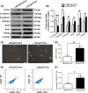
Pituitary tumor transforming gene1 (PTTG1) overexpression induced prostate cancer epithelial‐mesenchymal transition (EMT) and regulated the prostate cancer stem cell population. A,B, Western blotting demonstrated that PTTG1 overexpression resulted in down‐expression of E‐cadherin and up‐expression of N‐cadherin, vimentin, snail, Sox2 and Nanog, which were biomarkers of EMT and stem cells. (Data are presented as mean ± SD, *< .05 and **< .01.) C,D, Sphere formation assays were performed in LNCaP/PTTG1 and LNCaP/Control cells. LNCaP/PTTG1 cells formed greater sphere numbers than LNCaP/Control cells (Data are presented as mean ± SD, **< .01.) E,F, Using fluorescence activated cell sorting (FACS), prostate cancer stem cell population was quantified. CD44+/CD24 cell number in LNCaP/PTTG1 group was significantly increased compared with that in the LNCaP/Control group (Data are presented as mean ± SD, *< .05.)
Figure 5
Figure 5
Interleukin‐6 (IL‐6) increased pituitary tumor transforming gene 1 (PTTG1) expression via activating signal transducer and activator of transcription 3 (STAT3) in LNCaP cells. A‐D, IL‐6 was expressed in LNCaP cells with lentiviral transfection. After IL‐6 expression, PTTG1 protein and mRNA expressions were significantly increased in LNCaP/IL‐6 cells. 20 μM AG490 significantly weakened the effect of IL‐6 on PTTG1 expression. (Data are presented as mean ± SD, *< .05, **< .01 and ***< .001.) E, Schematic representation of potential STAT3 binding sites on PTTG1 promoter. F,G, Using luciferase reporter assays, the transcriptional activities of different PTTG1 promoter deletion constructs were quantified in LNCaP/Control and LNCaP/IL‐6 cells. (Data are presented as mean ± SD, ***< .001.) H, Putative STAT3 binding site sequence of PTTG1 were immunoprecipitated by an anti‐STAT3 antibody in extracts from LNCaP/IL‐6 cells. No signal was detected in the control IgG lane while control PCR reactions show robust amplification of the target sequence in input DNA
Figure 6
Figure 6
Signal transducer and activator of transcription 3 (STAT3) inhibitor, AG490, reversed bicalutamide resistance in DU145 cells. DU145 cells were treated with 5 μM bicalutamide combined with or without 20 μM AG490, clonogenic assays were performed and cell survival rate was calculated. As expected, we confirmed that DU145 cells were resistant to 5 μM bicalutamide treatment. However, the combined application with bicalutamide and AG490 significantly reduced the colony numbers (A and B) and cell survival rate (C) of DU145 cells compared with that of DU145 cells treated with single bicalutamide or AG490. (Data are presented as mean ± SD, **< .01 and ***< .001.)

Similar articles

Cited by

References

    1. Jemal A, Bray F, Center MM, Ferlay J, Ward E, Forman D. Global cancer statistics. CA Cancer J Clin. 2011;61:69‐90. - PubMed
    1. Ren SC, Chen R, Sun YH. Prostate cancer research in China. Asian J Androl. 2013;15:350‐353. - PMC - PubMed
    1. Karantanos T, Thompson TC. GEMMs shine a light on resistance to androgen deprivation therapy for prostate cancer. Cancer Cell. 2013;24:11‐13. - PMC - PubMed
    1. Patel LR, Barton MC. TRIM‐ing ligand dependence in castration‐resistant prostate cancer. Cancer Cell. 2016;29:776‐778. - PubMed
    1. Pei L, Melmed S. Isolation and characterization of a pituitary tumor‐transforming gene (PTTG). Mol Endocrinol. 1997;11:433‐441. - PubMed

MeSH terms