. 2018 Feb;129(2):360-367.
doi: 10.1016/j.clinph.2017.10.039. Epub 2017 Nov 16.

The effect of increased intracranial EEG sampling rates in clinical practice

Affiliations

The effect of increased intracranial EEG sampling rates in clinical practice

Kathryn A Davis et al. Clin Neurophysiol. 2018 Feb.

Abstract

Objective: Recent research suggests that high frequency intracranial EEG (iEEG) may improve localization of epileptic networks. This study aims to determine whether recording macroelectrode iEEG with higher sampling rates improves seizure localization in clinical practice.

Methods: 14 iEEG seizures from 10 patients recorded with >2000 Hz sampling rate were downsampled to four sampling rates: 100, 200, 500, 1000 Hz. In the 56 seizures, seizure onset time and location was marked by 5 independent, blinded EEG experts.

Results: When reading iEEG under clinical conditions, there was no consistent difference in time or localization of seizure onset or number of electrodes involved in the seizure onset zone with sampling rates varying from 100 to 1000 Hz. Stratification of patients by outcome did not improve with higher sampling rate.

Conclusion: When utilizing standard clinical protocols, there was no benefit to acquiring iEEGs with sampling rate >100 Hz. Significant variability was noted in EEG marking both within and between individual expert EEG readers.

Significance: Although commercial equipment is capable of sampling much faster than 100 Hz, tools allowing visualization of subtle high frequency activity such as HFOs will be required to improve patient care. Quantitative methods may decrease reader variability, and potentially improve patient outcomes.

Keywords: High frequency; Macroelectrode; Seizure onset.


Conflict of interest statement

Conflict of interest

None of the authors have potential conflicts of interest to be disclosed.

I confirm that I have read the Journal’s position on issues involved in ethical publication and affirm that this report is consistent with those guidelines.

Figures
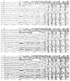
Fig. 1
Fig. 1
Comparison of EEG in patient 1 for 100 (top), 200 (middle), and 1000 (bottom) Hz sampling rate. The low voltage fast activity (red *) is clearly seen even at 200 Hz, and is visible as beta/gamma oscillations at 100 Hz. All five reviewers marked the onset at 2:32.2 seconds at all sampling rates. There was variability in the number of channels felt to be within the seizure onset zone, but no consistent correlation with sampling rate.
Figure 2
Figure 2
Comparison of EEG for patient 13 for 100 (top), 200 (middle) and 1000 (bottom) Hz sampling rate. There is a single broad area discharge (blue *), which at higher sampling rate is accompanied by low voltage fast activity, followed by a clear synchronous oscillations (red *) 6 seconds later. Reviewers picked between the two onset times, but were inconsistent.
Figure 3
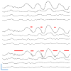
Figure 3. Visualization of HFOs
One second of 4 channels of data is shown from patient P002 during a seizure for 100 (top), 200 (middle) and 1000 Hz (bottom). Likely HFOs are shown with red bars. As expected, HFOs are poorly visualized by lower sampling rates. Although HFOs were visible during this seizure, no readers were able to identify interictal HFOs. Scale bar: 100 ms x 5 mV.

References

    1. Abend NS, Gutierrez-Colina A, Zhao H, Guo R, Marsh E, Clancy RR, et al. Interobserver reproducibility of electroencephalogram interpretation in critically ill children. J Clin Neurophysiol. 2011;28:15–9. doi: 10.1097/WNP.0b013e3182051123. - DOI - PMC - PubMed
    1. Benbadis SR, LaFrance WC, Papandonatos GD, Korabathina K, Lin K, Kraemer HC, et al. Interrater reliability of EEG-video monitoring. Neurology. 2009;73:843–6. doi: 10.1212/WNL.0b013e3181b78425. - DOI - PMC - PubMed
    1. Blanco JA, Stead M, Krieger A, Viventi J, Marsh WR, Lee KH, et al. Unsupervised Classification of High-Frequency Oscillations in Human Neocortical Epilepsy and Control Patients. J Neurophysiol. 2010;104:2900–12. doi: 10.1152/jn.01082.2009. - DOI - PMC - PubMed
    1. Cohen-Gadol AA, Wilhelmi BG, Collignon F, White JB, Britton JW, Cambier DM, et al. Long-term outcome of epilepsy surgery among 399 patients with nonlesional seizure foci including mesial temporal lobe sclerosis. J Neurosurg. 2006;104:513–24. doi: 10.3171/jns.2006.104.4.513. - DOI - PubMed
    1. Gardner AB, Worrell GA, Marsh E, Dlugos D, Litt B. Human and automated detection of high-frequency oscillations in clinical intracranial EEG recordings. Clin Neurophysiol. 2007;118:1134–43. doi: 10.1016/j.clinph.2006.12.019. - DOI - PMC - PubMed

Publication types