

Elevated expression of activated TH2 cells and milk-specific TH2 cells in milk-induced eosinophilic esophagitis
- PMID: 29289462
- PMCID: PMC5875940
- DOI: 10.1016/j.anai.2017.11.006
Elevated expression of activated TH2 cells and milk-specific TH2 cells in milk-induced eosinophilic esophagitis
Abstract
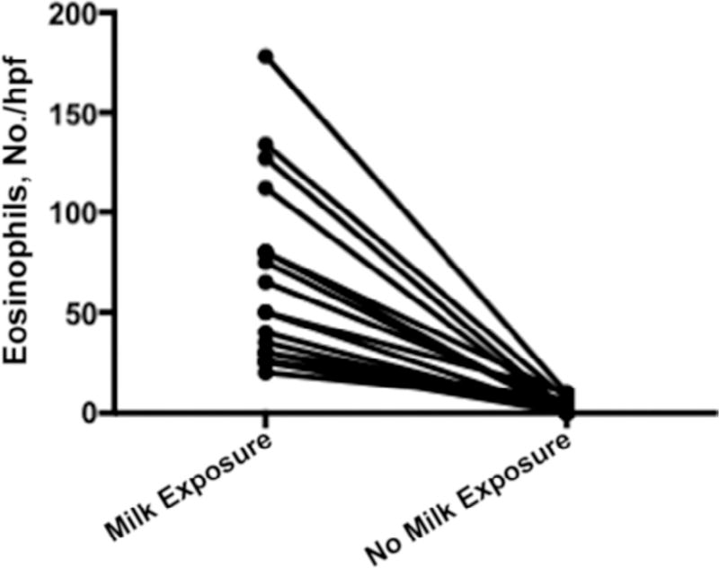
Background: Eosinophilic esophagitis (EoE) is an allergic inflammatory disease that is triggered by food allergens and characterized by progressive esophageal dysfunction. Esophageal biopsy specimens are characterized by eosinophilia and expression of TH2 cytokines.
Objective: To ascertain whether TH2 cells can exist in the peripheral blood in patients with milk-induced EoE.
Methods: Peripheral blood mononuclear cells from 20 children with milk-induced EoE were collected during active EoE (EoE-A) while consuming milk and inactive EoE (EoE-I) while not consuming milk, and 8 healthy patients without EoE were used as controls. The samples were analyzed for T-cell phenotype, including intracellular cytokines before and after incubation with milk antigens and assessed by flow cytometry.
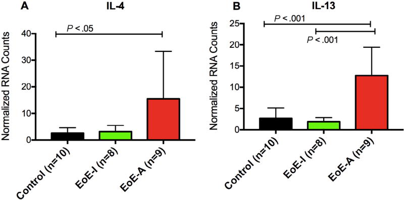
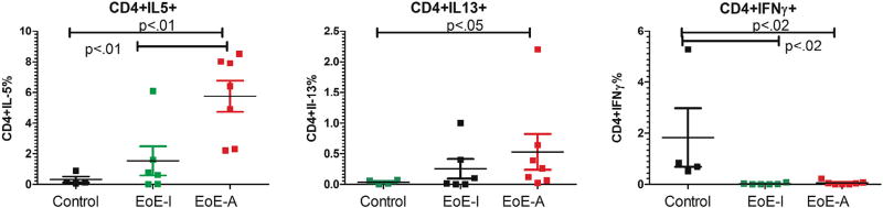
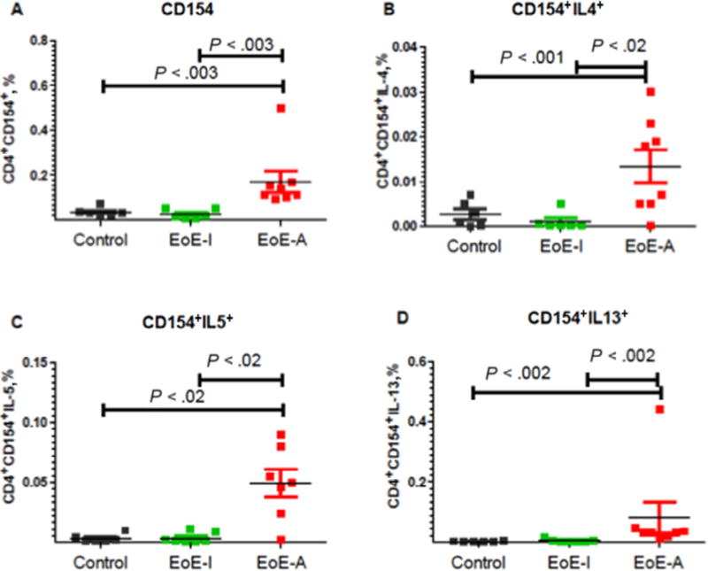
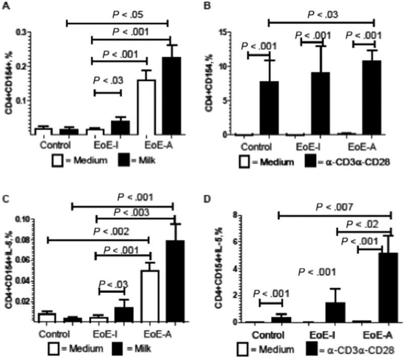
Results: We found a significant increase in CD4+ TH2 cells in the peripheral blood of patients with EoE-A compared with the controls. Furthermore, we observed a significant mean (SD) increase in the activation marker of CD154+ T cells (0.17% [0.047%]) in patients with EoE-A compared with control patients (0.034% [0.007%]) and EoE-I (0.025% [0.008]). These CD4+ T cells expressed significantly increase levels of TH2 cytokines (interleukins 4, 5, and 13) compared with the EoE-I and control groups. CD3+CD4+CD154+IL-5+ cells were significantly increased by milk antigens in both milk-induced EoE-A (0.050% [0.008%] to 0.079% [0.017%]) and EoE-I (0.0045% [0.002%] to 0.014% [0.008%]) compared with the controls (0.008% [0.003%] to 0.003% [0.001%]).
Conclusion: Our findings indicate that in EoE peripheral T cells have specific activation to milk allergens.
Copyright © 2017 American College of Allergy, Asthma & Immunology. Published by Elsevier Inc. All rights reserved.
Figures
Similar articles
-
Differential T follicular helper cell phenotypes distinguish IgE-mediated milk allergy from eosinophilic esophagitis in children.J Allergy Clin Immunol. 2025 Mar;155(3):909-922. doi: 10.1016/j.jaci.2024.09.024. Epub 2024 Oct 9. J Allergy Clin Immunol. 2025. PMID: 39389123
-
Invariant natural killer T cells in children with eosinophilic esophagitis.Clin Exp Allergy. 2014 Jan;44(1):58-68. doi: 10.1111/cea.12201. Clin Exp Allergy. 2014. PMID: 24118614 Free PMC article.
-
Allergic skin sensitization promotes eosinophilic esophagitis through the IL-33-basophil axis in mice.J Allergy Clin Immunol. 2016 Nov;138(5):1367-1380.e5. doi: 10.1016/j.jaci.2016.02.034. Epub 2016 Apr 19. J Allergy Clin Immunol. 2016. PMID: 27233150
-
Biological Therapies for Eosinophilic Esophagitis: Where Do We Stand?Clin Rev Allergy Immunol. 2018 Oct;55(2):205-216. doi: 10.1007/s12016-018-8674-3. Clin Rev Allergy Immunol. 2018. PMID: 29372536 Review.
-
Eosinophilic Esophagitis and Gastroenteritis.Curr Allergy Asthma Rep. 2015 Sep;15(9):58. doi: 10.1007/s11882-015-0558-5. Curr Allergy Asthma Rep. 2015. PMID: 26233430 Review.
Cited by
-
Allergies and Eosinophilic Esophagitis-Current Updates for the Pediatric Gastroenterologist.Curr Gastroenterol Rep. 2019 Nov 20;21(11):56. doi: 10.1007/s11894-019-0729-y. Curr Gastroenterol Rep. 2019. PMID: 31748971 Free PMC article. Review.
-
Viral Induced Effects on a Vulnerable Epithelium; Lessons Learned From Paediatric Asthma and Eosinophilic Oesophagitis.Front Immunol. 2021 Nov 29;12:773600. doi: 10.3389/fimmu.2021.773600. eCollection 2021. Front Immunol. 2021. PMID: 34912343 Free PMC article. Review.
-
Expression profiling identifies key genes and biological functions associated with eosinophilic esophagitis in human patients.Front Allergy. 2023 Aug 24;4:1239273. doi: 10.3389/falgy.2023.1239273. eCollection 2023. Front Allergy. 2023. PMID: 37692891 Free PMC article.
-
IL-13-induced STAT3-dependent signaling networks regulate esophageal epithelial proliferation in eosinophilic esophagitis.J Allergy Clin Immunol. 2023 Dec;152(6):1550-1568. doi: 10.1016/j.jaci.2023.07.021. Epub 2023 Aug 29. J Allergy Clin Immunol. 2023. PMID: 37652141 Free PMC article.
-
Differential T follicular helper cell phenotypes distinguish IgE-mediated milk allergy from eosinophilic esophagitis in children.J Allergy Clin Immunol. 2025 Mar;155(3):909-922. doi: 10.1016/j.jaci.2024.09.024. Epub 2024 Oct 9. J Allergy Clin Immunol. 2025. PMID: 39389123
References
-
- Liacouras CA, Furuta GT, Hirano I, et al. Eosinophilic esophagitis: updated consensus recommendations for children and adults. J Allergy Clin Immunol. 2011;128:3–20. e26. quiz 21–22. - PubMed
Publication types
MeSH terms
Substances
Grants and funding
LinkOut - more resources
Full Text Sources
Other Literature Sources
Medical
Research Materials
