. 2018 Feb:8:1-12.
doi: 10.1016/j.molmet.2017.12.006. Epub 2017 Dec 18.

Differential contribution of POMC and AgRP neurons to the regulation of regional autonomic nerve activity by leptin

Affiliations

Differential contribution of POMC and AgRP neurons to the regulation of regional autonomic nerve activity by leptin

Balyssa B Bell et al. Mol Metab. 2018 Feb.

Erratum in

Abstract

Objectives: The autonomic nervous system is critically involved in mediating the control by leptin of many physiological processes. Here, we examined the role of the leptin receptor (LepR) in proopiomelanocortin (POMC) and agouti-related peptide (AgRP) neurons in mediating the effects of leptin on regional sympathetic and parasympathetic nerve activity.

Methods: We analyzed how deletion of the LepR in POMC neurons (POMCCre/LepRfl/fl mice) or AgRP neurons (AgRPCre/LepRfl/fl mice) affects the ability of leptin to increase sympathetic and parasympathetic nerve activity. We also studied mice lacking the catalytic p110α or p110β subunits of phosphatidylinositol-3 kinase (PI3K) in POMC neurons.

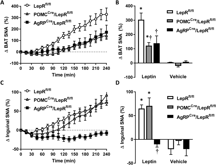
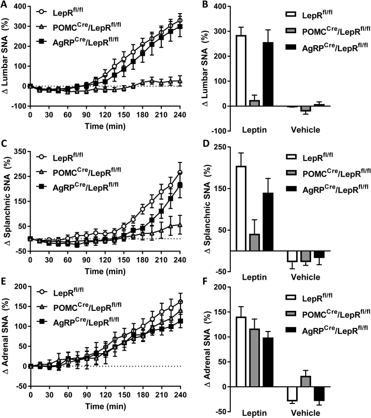
Results: Leptin-evoked increase in sympathetic nerve activity subserving thermogenic brown adipose tissue was partially blunted in mice lacking the LepR in either POMC or AgRP neurons. On the other hand, loss of the LepR in AgRP, but not POMC, neurons interfered with leptin-induced sympathetic nerve activation to the inguinal fat depot. The increase in hepatic sympathetic traffic induced by leptin was also reduced in mice lacking the LepR in AgRP, but not POMC, neurons whereas LepR deletion in either AgRP or POMC neurons attenuated the hepatic parasympathetic nerve activation evoked by leptin. Interestingly, the renal, lumbar and splanchnic sympathetic nerve activation caused by leptin were significantly blunted in POMCCre/LepRfl/fl mice, but not in AgRPCre/LepRfl/fl mice. However, loss of the LepR in POMC or AgRP neurons did not interfere with the ability of leptin to increase sympathetic traffic to the adrenal gland. Furthermore, ablation of the p110α, but not the p110β, isoform of PI3K from POMC neurons eliminated the leptin-elicited renal sympathetic nerve activation. Finally, we show trans-synaptic retrograde tracing of both POMC and AgRP neurons from the kidneys.

Conclusions: POMC and AgRP neurons are differentially involved in mediating the effects of leptin on autonomic nerve activity subserving various tissues and organs.

Keywords: Autonomic nervous system; Cardiovascular regulation; Energy homeostasis; Leptin.


Figures

None
Graphical abstract
Figure 1
Figure 1
LepR deletion from POMC or AgRP neurons prevents leptin-induced STAT3 phosphorylation. A-B) In control mice, ICV leptin treatment increased STAT3 phosphorylation in both POMC (A) and AgRP (B) neurons. C-D) Loss of LepR from POMC (C) or AgRP (D) neurons abolished leptin-induced phosphorylation of STAT3 in those neurons. Images acquired at 20× magnification. Scale bar, 100 μm. E-F) Relative to littermate controls, male and female mice lacking the LepR in POMC (E, n = 7–19 male, 9–23 female LepRfl/fl; n = 16–35 male, 10–27 female POMCCre/LepRfl/fl) or AgRP (F, n = 7–9 male, 9–10 female LepRfl/fl; n = 12–21 male, 13–17 female AgRPCre/LepRfl/fl) neurons display increased body weight.
Figure 2
Figure 2
Contrasting effects of LepR ablation in POMC and AgRP neurons on leptin control of BAT and WAT SNA. A–B) Leptin-induced increase in BAT SNA is partially attenuated in both POMCCre/LepRfl/fl and AgRPCre/LepRfl/fl mice (n = 11 leptin treated LepRfl/fl mice; n = 9 leptin treated POMCCre/LepRfl/fl mice; n = 9 leptin treated AgRPCre/LepRfl/fl mice; n = 9 vehicle treated LepRfl/fl mice; n = 7 vehicle treated POMCCre/LepRfl/fl mice; n = 5 vehicle treated AgRPCre/LepRfl/fl mice). C–D) Sympathetic nerve activation to inguinal WAT elicited by ICV leptin is impaired in AgRPCre/LepRfl/fl mice, but not POMCCre/LepRfl/fl mice (n = 15 leptin treated LepRfl/fl mice; n = 13 leptin treated POMCCre/LepRfl/fl mice; n = 17 leptin treated AgRPCre/LepRfl/fl mice; n = 5 vehicle treated LepRfl/fl mice; n = 6 vehicle treated POMCCre/LepRfl/fl mice; n = 6 vehicle treated AgRPCre/LepRfl/fl mice). Bar graphs represent the averages of the last hour of SNA recordings. Comparisons were made using 2-way ANOVA with or without repeated measures, *p < 0.05 vs genotype matched vehicle, p < 0.05 vs leptin-LepRfl/fl controls.
Figure 3
Figure 3
Loss of LepR in POMC and AgRP neurons affects differentially the effects of leptin on hepatic autonomic nerve activity. A–B) Leptin stimulation of hepatic SNA was partially blunted in AgRPCre/LepRfl/fl mice, but not POMCCre/LepRfl/fl mice (n = 12 leptin treated LepRfl/fl mice; n = 9 leptin treated POMCCre/LepRfl/fl mice; n = 6 leptin treated AgRPCre/LepRfl/fl mice; n = 3 vehicle treated LepRfl/fl mice; n = 3 vehicle treated POMCCre/LepRfl/fl mice; n = 9 vehicle treated AgRPCre/LepRfl/fl mice). C–D) The increase in hepatic PSNA evoked by leptin was abolished in both POMCCre/LepRfl/fl and AgRPCre/LepRfl/fl mice (n = 15 leptin treated LepRfl/fl mice; n = 13 leptin treated POMCCre/LepRfl/fl mice; n = 17 leptin treated AgRPCre/LepRfl/fl mice; n = 5 vehicle treated LepRfl/fl mice; n = 6 vehicle treated POMCCre/LepRfl/fl mice; n = 6 vehicle treated AgRPCre/LepRfl/fl mice). Bar graphs represent the averages of the last hour of SNA and PNSA recordings. Comparisons were made using 2-way ANOVA with or without repeated measures, *p < 0.05 vs genotype matched vehicle, p < 0.05 vs leptin-LepRfl/fl controls.
Figure 4
Figure 4
The LepR on POMC, but not AgRP, neurons is required for leptin activation of lumbar and splanchnic SNA, but not adrenal SNA. A–D) ICV leptin (2 μg) increased lumbar (A-B, n = 13 leptin treated LepRfl/fl mice; n = 8 leptin treated POMCCre/LepRfl/fl mice; n = 8 leptin treated AgRPCre/LepRfl/fl mice; n = 5 vehicle treated LepRfl/fl mice; n = 6 vehicle treated POMCCre/LepRfl/fl mice; n = 5 vehicle treated AgRPCre/LepRfl/fl mice), and splanchnic (C-D, n = 12 leptin treated LepRfl/fl mice; n = 9 leptin treated POMCCre/LepRfl/fl mice; n = 6 leptin treated AgRPCre/LepRfl/fl mice; n = 3 vehicle treated LepRfl/fl mice; n = 4 vehicle treated POMCCre/LepRfl/fl mice; n = 5 vehicle treated AgRPCre/LepRfl/fl mice) SNA in control and AgRPCre/LepRfl/fl mice; but not in POMCCre/LepRfl/fl mice. E–F) The leptin-evoked increase in SNA subserving the adrenal gland was not affected in POMCCre/LepRfl/fl and AgRPCre/LepRfl/fl mice (n = 14 leptin treated LepRfl/fl mice; n = 9 leptin treated POMCCre/LepRfl/fl mice; n = 7 leptin treated AgRPCre/LepRfl/fl mice; n = 3 vehicle treated LepRfl/fl mice; n = 3 vehicle treated POMCCre/LepRfl/fl mice; n = 3 vehicle treated AgRPCre/LepRfl/fl mice). Bar graphs represent the averages of the last hour of SNA recordings. Comparisons were made using 2-way ANOVA with repeated or without measures, *p < 0.05 vs genotype matched vehicle, p < 0.05 vs leptin-LepRfl/fl controls.
Figure 5
Figure 5
LepR ablation from POMC, but not AgRP, neurons effects selectively leptin control of renal SNA. A–C) Leptin administered ICV (2 μg, A-B, n = 13 leptin treated LepRfl/fl mice; n = 8 leptin treated POMCCre/LepRfl/fl mice; n = 8 leptin treated AgRPCre/LepRfl/fl mice; n = 5 vehicle treated LepRfl/fl mice; n = 6 vehicle treated POMCCre/LepRfl/fl mice; n = 5 vehicle treated AgRPCre/LepRfl/fl mice) or intravenously (120 μg, C, n = 16 LepRfl/fl mice; n = 8 POMCCre/LepRfl/fl mice; n = 8 AgRPCre/LepRfl/fl mice) increased renal SNA in control (LepRfl/fl) and AgRPCre/LepRfl/fl mice, but not in POMCCre/LepRfl/fl mice. D) POMCCre/LepRfl/fl mice displayed a normal increase in renal SNA in response to ICV insulin (n = 6 LepRfl/fl mice; n = 8 POMCCre/LepRfl/fl mice). The bar graph represents the averages of the last hour of SNA recordings. Comparisons were made using 2-way ANOVA with or without repeated measures, *p < 0.05 vs genotype matched vehicle, p < 0.05 vs leptin-LepRfl/fl controls.
Figure 6
Figure 6
Effects of POMC neurons-selective deletion of p110α or p110β catalytic subunits of PI3K. A–B) Deletion of the p110α (A) or p110β (B) had no effect on body weight in male or female mice fed normal chow or male mice fed a high fat diet (HFD). C–D) Leptin-induced increase in renal SNA, measured in the conscious state, was abolished in mice lacking the p110α, but not p110β, PI3K in POMC neurons (n = 9 p110αfl/fl + p110 βfl/fl mice; n = 6 POMCCre/p110αfl/fl mice; n = 6 p110 βfl/fl mice). The bar graph represents the averages of the last hour of SNA recordings. Comparisons were made using t-test, 1- or 2-way ANOVA with or without repeated measures, *p < 0.05 vs p110αfl/fl + p110 βfl/fl mice.
Figure 7
Figure 7
Trans-synaptic retrograde tracing of POMC and AgRP neurons from the kidneys. A–B) GFP-expressing PRV-152 pseudorabies virus injected into the left and right kidneys trans-synaptically labels, after 5 days, the ARC POMC (A) and AgRP neurons (B) in POMCCre/tdTomato mice and AgRPCre/tdTomato mice, respectively. Images acquired at 20× magnification. Scale bar, 100 μm.

Comment in

References

    1. Friedman J.M., Mantzoros C.S. 20 years of leptin: from the discovery of the leptin gene to leptin in our therapeutic armamentarium. Metabolism. 2015;64:1–4. - PubMed
    1. Cui H., Lopez M., Rahmouni K. The cellular and molecular bases of leptin and ghrelin resistance in obesity. Nature Reviews Endocrinology. 2017;13:338–351. - PMC - PubMed
    1. Plum L., Rother E., Munzberg H., Wunderlich F.T., Morgan D.A., Hampel B. Enhanced leptin-stimulated Pi3k activation in the CNS promotes white adipose tissue transdifferentiation. Cell Metabolism. 2007;6:431–445. - PubMed
    1. Buettner C., Muse E.D., Cheng A., Chen L., Scherer T., Pocai A. Leptin controls adipose tissue lipogenesis via central, STAT3-independent mechanisms. Nature Medicine. 2008;14:667–675. - PMC - PubMed
    1. Scherer T., Buettner C. Yin and Yang of hypothalamic insulin and leptin signaling in regulating white adipose tissue metabolism. Reviews in Endocrine & Metabolic Disorders. 2011;12:235–243. - PMC - PubMed

Publication types

MeSH terms