. 2018 Jan 4;102(1):116-132.
doi: 10.1016/j.ajhg.2017.12.002. Epub 2017 Dec 28.

KIAA1109 Variants Are Associated with a Severe Disorder of Brain Development and Arthrogryposis

Affiliations

KIAA1109 Variants Are Associated with a Severe Disorder of Brain Development and Arthrogryposis

Lucie Gueneau et al. Am J Hum Genet. .

Abstract

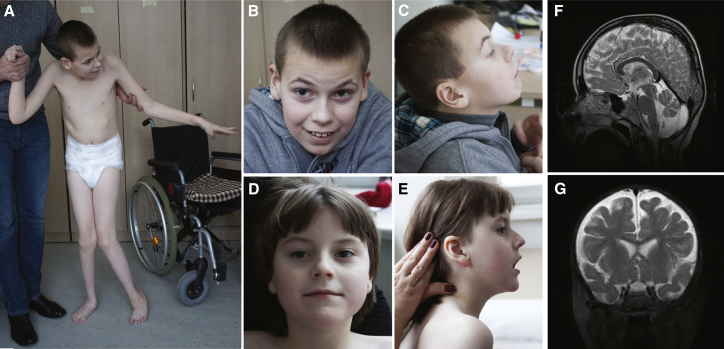
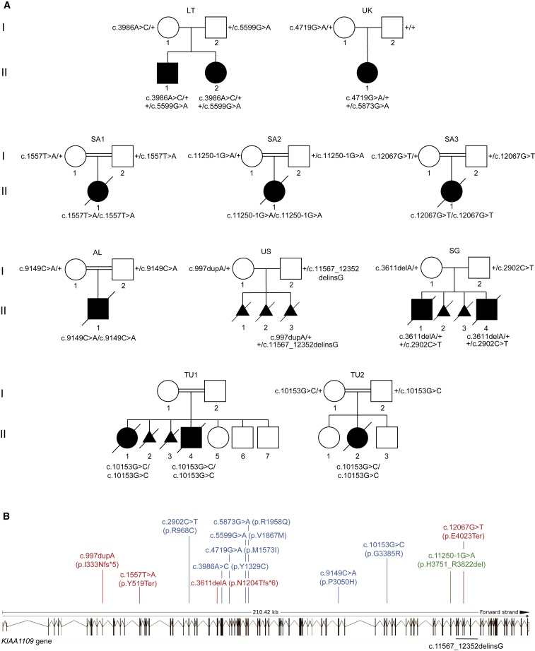
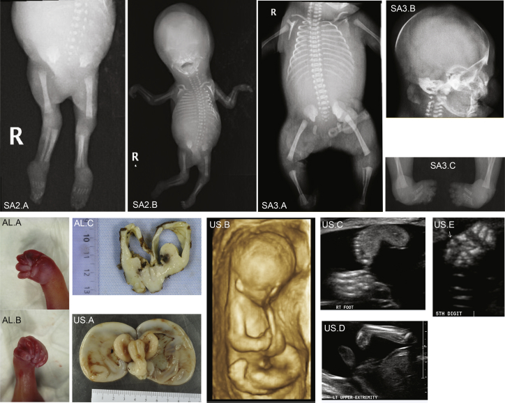
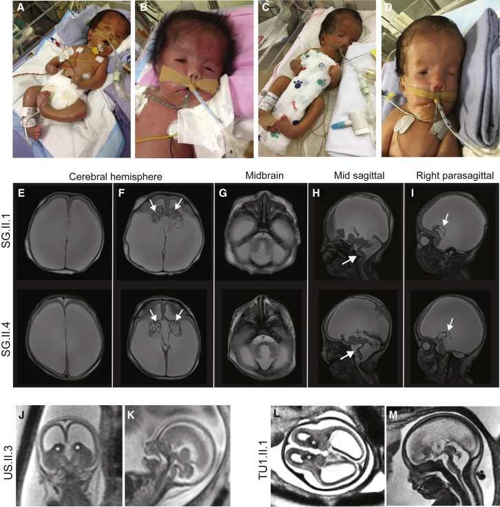
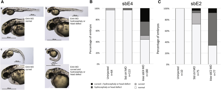
Whole-exome and targeted sequencing of 13 individuals from 10 unrelated families with overlapping clinical manifestations identified loss-of-function and missense variants in KIAA1109 allowing delineation of an autosomal-recessive multi-system syndrome, which we suggest to name Alkuraya-Kučinskas syndrome (MIM 617822). Shared phenotypic features representing the cardinal characteristics of this syndrome combine brain atrophy with clubfoot and arthrogryposis. Affected individuals present with cerebral parenchymal underdevelopment, ranging from major cerebral parenchymal thinning with lissencephalic aspect to moderate parenchymal rarefaction, severe to mild ventriculomegaly, cerebellar hypoplasia with brainstem dysgenesis, and cardiac and ophthalmologic anomalies, such as microphthalmia and cataract. Severe loss-of-function cases were incompatible with life, whereas those individuals with milder missense variants presented with severe global developmental delay, syndactyly of 2nd and 3rd toes, and severe muscle hypotonia resulting in incapacity to stand without support. Consistent with a causative role for KIAA1109 loss-of-function/hypomorphic variants in this syndrome, knockdowns of the zebrafish orthologous gene resulted in embryos with hydrocephaly and abnormally curved notochords and overall body shape, whereas published knockouts of the fruit fly and mouse orthologous genes resulted in lethality or severe neurological defects reminiscent of the probands' features.

Keywords: arthrogryposis; brain malformations; cerebellar hypoplasia; clubfoot; hydrocephaly; whole-exome sequencing.


Figures

Figure 1
Figure 1
Pictures and Brain MRI from Surviving Individuals Front and side views of the LT affected brother LT.II.1 (A–C) and sister LT.II.2 (D, E) at the ages of 13 years and 7 years, respectively. Brain MRI images of affected individual LT.II.1 at age of 8 years showed small posterior fossa arachnoid cyst, discrete vermian atrophy, and slight increase of the fluid-filled retro and infra-cerebellar space (F). Brain MRI images of affected individual LT.II.2 at age of 1 year showed discrete parenchymal rarefaction involving mainly the frontal lobes (G).
Figure 2
Figure 2
KIAA1109 Pedigrees and Variants (A) Pedigrees of the ten families carrying KIAA1109 variants. The affected individuals of the Lithuanian (LT), Singaporean (SG), British (UK), and American (US) families are compound heterozygotes for rare variants, whereas the probands of the Algerian (AL), Saudi Arabian (SA1–SA3), and Tunisian (TU1, TU2) consanguineous families are homozygous for KIAA1109 variants. (B) Distribution of variants along the schematically represented 86 exons of KIAA1109. Missense variants are depicted in blue, nonsense in red, and the splice site variant in green. The extent of the deletion identified in the proband of the US family is indicated in black below.
Figure 3
Figure 3
Ultrasound, X-Rays, and Autopsy Images of the SA2.II.1, SA3.II.1, AL.II.1, and US.II.3 Fetuses X-ray images showing arthrogryposis of SA2.II.1 fetus (SA2.A and SA2.B). X-ray images showing SA3.II.1 skeleton (SA3.A), head (SA3.B), and club feet (SA3.C). Autopsy pictures from the AL.II.1 fetus showing right (AL.A) and left (AL.B) adductus thumbs of the fetus, and dilatation of cerebral ventricles with agenesis of corpus callosum (AL.C). Autopsy image of the brain from US.II.3 fetus showing hydrocephalic brain with diaphanous pallium (US.A). The colliculi appear as single elongated ridges separated by a midline futter and the midline appears angulated on the brainstem, which is small as is the cerebellum. Antenatal ultrasound scan showed general arthrogryposis (US.B), one hyperflexed wrist (US.C), club feet (US.D), and bilateral clinodactyly of one hand (US.E).
Figure 4
Figure 4
Pictures and Brain MRI Images of the SG.II.1 and SG.II.4 Babies and US.II.3 and TU1.II.1 Fetuses (A–D) Photographs of SG.II.1 (A and B) and SG.II.4 (C and D) babies showing their whole bodies (A and C) and a close up of their faces (B and D). (E–I) Brain MRI images of the elder brother SG.II.1 (top) and the younger brother SG.II.4 (bottom). Axial T2 weighted images showed severe ventriculomegaly, associated with severe thinning of the brain parenchyma (E, F). The brain parenchyma showed absence of normal gyral/sulcal pattern with smooth appearance in keeping with lissencephaly (E, F). Corpus callosum appeared to be absent (E, H). Note the prominent germinal matrix with germinolysis cysts (solid arrows) (F, I). The pons and cerebellum appeared hypoplastic with dilatation of the 4th ventricle (G, H) and Z shaped appearance of the brainstem (solid arrows) (H). (J–M) Coronal (J), axial (L), and midsagittal (K, M) T2-weighed fetal prenatal MRI images of US.II.3 at 18.5 weeks of pregnancy (J and K) and TU1.II.1 at 28 weeks of pregnancy (L and M) demonstrating a similar imaging pattern including thin parenchyma (lissencephalic aspect), prominent germinal matrix marked by an asterisk, ventriculomegaly, and brain stem and vermian dysgenesis (kinked brain stem and elongated pons). In summary, we observe a similar brain malformation pattern both prenatally—US.II.3 in (J) and (K), TU1.II.1 in (L) and (M), AL.II.1 (see text), TU1.II.4 (see text), and TU2.II.2 (see text)—and postnatally (SG.II.1 [E–I top] and SG.II.4 [E–I bottom]).
Figure 5
Figure 5
kiaa1109 Knockdown in Zebrafish Results in Phenotypes Reminiscent of Probands’ Clinical Features (A) Lateral views representing the four classes of observed phenotypes in 2 dpf TU zebrafish embryos injected with sbE4-MO (morpholino) targeting kiaa1109: from top left to bottom right, normal, hydrocephalic or other head defects, curved and curved with head defect. (B) Results for uninjected embryos (left) and those injected with equivalent amounts of standard control MO (center) or kiaa1109 sbE4-MO (6.7 ng, right). Phenotyping and scoring were performed at 2 dpf in two independent experiments. (C) Results for uninjected embryos (left) and those injected with equivalent amounts of standard control MO (center) or kiaa1109 sbE2-MO (16.9 ng, right). Phenotyping and scoring were performed at 2 dpf in two independent experiments.

References

    1. Yang Y., Muzny D.M., Reid J.G., Bainbridge M.N., Willis A., Ward P.A., Braxton A., Beuten J., Xia F., Niu Z. Clinical whole-exome sequencing for the diagnosis of mendelian disorders. N. Engl. J. Med. 2013;369:1502–1511. - PMC - PubMed
    1. Anazi S., Maddirevula S., Faqeih E., Alsedairy H., Alzahrani F., Shamseldin H.E., Patel N., Hashem M., Ibrahim N., Abdulwahab F. Clinical genomics expands the morbid genome of intellectual disability and offers a high diagnostic yield. Mol. Psychiatry. 2017;22:615–624. - PubMed
    1. Vissers L.E., Gilissen C., Veltman J.A. Genetic studies in intellectual disability and related disorders. Nat. Rev. Genet. 2016;17:9–18. - PubMed
    1. Mirzaa G.M., Paciorkowski A.R. Introduction: Brain malformations. Am. J. Med. Genet. C. Semin. Med. Genet. 2014;166C:117–123. - PubMed
    1. Kato M. Genotype-phenotype correlation in neuronal migration disorders and cortical dysplasias. Front. Neurosci. 2015;9:181. - PMC - PubMed

Publication types

LinkOut - more resources