. 2018 Jan 1;8(1):199-211.
doi: 10.7150/thno.21425. eCollection 2018.

Histidine-rich Modification of a Scorpion-derived Peptide Improves Bioavailability and Inhibitory Activity against HSV-1

Affiliations

Histidine-rich Modification of a Scorpion-derived Peptide Improves Bioavailability and Inhibitory Activity against HSV-1

Zhengyang Zeng et al. Theranostics. .

Abstract

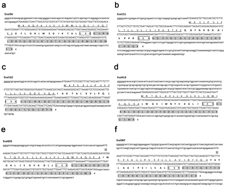
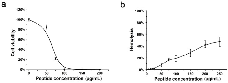
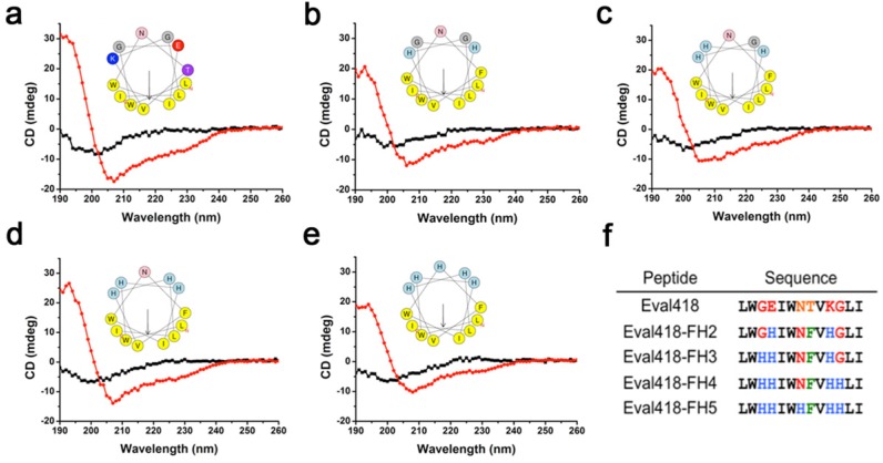
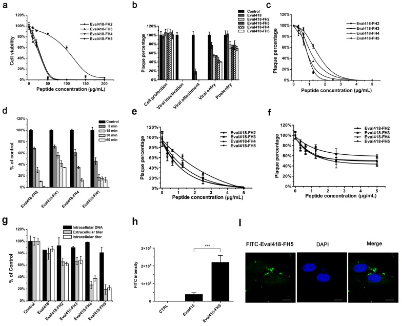
Rationale: HSV is one of the most widespread human viral pathogens. HSV-1 infects a large portion of the human population and causes severe diseases. The current clinical treatment for HSV-1 is based on nucleoside analogues, the use of which is limited due to drug resistance, side effects and poor bioavailability. AMPs have been identified as potential antiviral agents that may overcome these limitations. Therefore, we screened anti-HSV-1 peptides from a scorpion-derived AMP library and engineered one candidate into a histidine-rich peptide with significantly improved antiviral activity and development potential. Methods: A venomous gland cDNA library was constructed from the scorpion Euscorpiops validus in the Yunnan Province of China. Six putative AMPs were characterized from this cDNA library, and the synthesized peptides were screened via plaque-forming assays to determine their virucidal potential. Time of addition experiments according to the infection progress of HSV-1 were used to identify the modes of action for peptides of interest. The histidine-rich modification was designed based on structural analysis of peptides by a helical wheel model and CD spectroscopy. Peptide cellular uptake and distribution were measured by flow cytometry and confocal microscopy, respectively. Results: The peptide Eval418 was found to have high clearance activity in an HSV-1 plaque reduction assay. Eval418 exhibited dose-dependent and time-dependent inactivation of HSV-1 and dose-dependent inhibition of HSV-1 attachment to host cells. However, Eval418 scarcely suppressed an established HSV-1 infection due to poor cellular uptake. We further designed and modified Eval418 into four histidine-rich derivative peptides with enhanced antiviral activities and lower cytotoxicities. All of the derivative peptides suppressed established HSV-1 infections. One of these peptides, Eval418-FH5, not only had strong viral inactivation activity and enhanced attachment inhibitory activity but also had high inhibitory activity against intracellular HSV-1, which was consistent with its improved intracellular uptake and distribution as confirmed by confocal microscopy and flow cytometry. Conclusion: We successfully identified an anti-HSV-1 peptide, Eval418, from a scorpion venom peptide library and designed a histidine-rich Eval418 derivative with significantly improved potential for further development as an anti-HSV-1 drug. This successful modification can provide a design strategy to improve the bioavailability, cellular distribution and antiviral activity of peptide agents.

Keywords: Anti-HSV-1 peptides; Bioavailability.; Cellular uptake; Histidine-rich modification; Scorpion venom peptides.


Conflict of interest statement

Competing Interests: The authors have declared that no competing interest exists.

Figures

Figure 1
Figure 1
Precursor structure analysis of six scorpion-derived peptides. Six scorpion peptides from the venomous cDNA library of the scorpion E. validus were characterized as candidate antimicrobial agents, and their precursor structures were analyzed. The amino acid sequences from open-reading frames are shown. The signal peptide and pro-peptide regions are underlined and highlighted with a gray background, respectively. The cutting signals are shown in frames, and the mature peptide regions are bolded.
Figure 2
Figure 2
Screening of anti-HSV-1 peptides from the venomous cDNA library of the scorpion E. validus. The six scorpion peptides were prepared by chemical synthesis and screened for their abilities to inactivate HSV-1. (a) The inhibitory activities of the six candidate peptides at 10 μg/mL against HSV-1 proliferation were measured by plaque reduction assay in Vero cells. (b) Comparison of inhibitory rates of the six scorpion peptides against HSV-1 proliferation. The plaques in each well were counted, and the inhibitory rates were calculated. The values represent the mean ± SEM of five independent samples. The drug ACV (10 μg/mL) was used as a positive control. ***P< 0.001. (c) Sequence alignments of the six scorpion peptides. The sequence alignments of the candidate antimicrobial peptides were performed using Genedoc software. Hydrophilic and hydrophobic residues are marked by colorful and colorless backgrounds, respectively.
Figure 3
Figure 3
Cytotoxicity and hemolytic activity of the Eval418 peptide. (a) Cytotoxicity of Eval418 peptide was measured on Vero cells by MTT assay. The concentrations ranged from 0 to 200 μg/mL. The 50% cytotoxicity concentration (CC50) of Eval418 to Vero cells was 68.50 μg/mL. (b) The hemolytic activity of Eval418 was evaluated in human erythrocytes by hemolytic assay. The concentrations also ranged from 0 to 200 μg/mL. The hemolysis rate of human erythrocytes was less than 50% when the concentration of Eval418 was as high as 200 μg/mL.
Figure 4
Figure 4
Comprehensive anti-HSV-1 activities and cellular localization of the Eval418 peptide. (a) Time of addition experiments were conducted to determine the anti-HSV-1 activity of the Eval418 peptide. The upper panel is a schematic overview of the operational approach for the time of addition experiments. The cells or viruses were treated with Eval418 peptide at a final concentration of 10 μg/mL under five diverse treatment modes. The lower panel shows the anti-HSV-1 activity assays of Eval418 in the time of addition experiments. The inhibitory effects of Eval418 in each treatment mode were determined by plaque reduction assay. ***P< 0.001. (b) Concentration-dependent inactivation activity of the Eval418 peptide. HSV-1 (60 PFU/well) was directly incubated with the Eval418 peptide. The virus-peptide mixture was then diluted to the indicated concentrations and applied to plaque reduction assay on Vero cells. (c) Time-dependent inactivation activity of the Eval418 peptide. HSV-1 (60 PFU/well) was directly incubated with the Eval418 peptide over serial durations, and the mixture was applied to a plaque reduction assay on Vero cells. (d) Inhibitory activity of the Eval418 peptide against HSV-1 viral attachment. Eval418 was added to the cell culture medium at a series of concentrations simultaneously with HSV-1 at the viral attachment step and rinsed out before viral entry. Then, the inhibitory effect was measured by plaque reduction assay. (e) The inhibitory activity of the Eval418 peptide against HSV-1 at the viral entry step. After viral attachment, Eval418 was added and co-incubated with cells during the viral entry step. The inhibitory effect was then measured by plaque reduction assay. (f) Cellular localization of the Eval418 peptide in Vero cells. Eval418 was labeled by FITC, and the cellular localization of Eval418 was determined by confocal microscopy after incubation for 24 h with Vero cells. Scale bars: 10 µM.
Figure 5
Figure 5
Design of Eval418 peptide derivatives. The CD spectra measurements were performed over a UV range of 250-190 nm at 25 ℃ in water on a jasco-810 spectropolarimeter (a-e). The helical wheels showed the hydrophilic and hydrophobic faces of Eval418 (a) and its derivative peptides, which were designated Eval418-FH2 (b), Eval418-FH3 (c), Eval418-FH4 (d) and Eval418-FH5 (e). (f) Sequence alignments of the Eval418 peptide and its derivative peptides.
Figure 6
Figure 6
Cytotoxicities, anti-HSV-1 activities and intracellular distributions of the Eval418-derived peptides. (a) Cytotoxicities of Eval418-derived peptides. The cytotoxicities of Eval418-FH2, Eval418-FH3, Eval418-FH4 and Eval418-FH5 were measured on Vero cells by MTT assay. The concentrations ranged from 0 to 200 μg/mL, and the CC50 values were 27.60, 26.83, 27.58 and 106.68 μg/mL, respectively. (b) Anti-HSV-1 activity assay of the Eval418-derived peptides in the time of addition experiment. The inhibitory effects of the Eval418-derived peptides in each treatment mode were determined by plaque reduction assay. (c) Concentration-dependent inactivation activities of Eval418 derivative peptides. HSV-1 (60 PFU/well) was directly treated with Eval418-derived peptides. Vero cells were infected with the virus-peptide mixtures at a serial final concentration of the derivative peptides. (d) Time-dependent inactivation activities of Eval418 derivative peptides. Eval418-derived peptides were added to HSV-1 (60 PFU/well) and incubated over a series of indicated durations. The mixtures were then used to infect Vero cells, and the plaque reduction assay was completed. (e) Inhibitory activities of the Eval418-derived peptides when added to cells during the HSV-1 viral attachment step. Peptides were added to culture medium of Vero cells simultaneously with HSV-1 and incubated for 1 h at 4 ℃. The cells were then rinsed and replenished with a cover layer, and the plaque reduction assay was completed. (f) Inhibitory activities of the Eval418-derived peptides against viral entry of HSV-1. Cells were incubated with HSV-1 at 4 ℃ for 1 h. After HSV-1 was washed out, the derivative peptides were added and incubated at 37 ℃ for 1 h. The cells were then rinsed and replenished with a cover layer. The inhibitory rates were measured by plaque reduction assay. (g) Anti-HSV-1 activities of Eval418 and its derivative peptides during the post-entry step. Extracellular and intracellular HSV-1 infectivity levels were assessed by plaque forming assay, and the DNA content was assessed by real-time PCR. (h) Flow cytometry measurement of the cellular uptake of Eval418 and Eval418-FH5 peptides. Vero cells were incubated with FITC-labeled Eval418 or Eval418-FH5 for 24 h, and the average FITC intensity of each cell was measured by flow cytometry. ***P< 0.001. (i) Confocal microscopic examination of cellular localization of Eval418-FH5. Eval418-FH5 was labeled by FITC and used to treat Vero cells for 24 h. The cellular localization was determined using confocal microscopy. Scale bars: 10 µM.

Similar articles

Cited by

References

    1. Brandt C R. The role of viral and host genes in corneal infection with herpes simplex virus type 1. Experimental eye research. 2005;80:607–21. - PubMed
    1. Liashkovich I, Hafezi W, Kuhn J M, Oberleithner H, Shahin V. Nuclear delivery mechanism of herpes simplex virus type 1 genome. Journal of molecular recognition: JMR. 2011;24:414–21. - PubMed
    1. Schacker T. The role of HSV in the transmission and progression of HIV. Herpes: the journal of the IHMF. 2001;8:46–9. - PubMed
    1. Schang L M. Herpes simplex viruses in antiviral drug discovery. Current pharmaceutical design. 2006;12:1357–70. - PubMed
    1. Diamond G, Beckloff N, Weinberg A, Kisich K O. The roles of antimicrobial peptides in innate host defense. Current pharmaceutical design. 2009;15:2377–92. - PMC - PubMed

Publication types

MeSH terms

LinkOut - more resources