Review
. 2018 Jan 1;8(1):237-255.
doi: 10.7150/thno.21945. eCollection 2018.

Exosome Theranostics: Biology and Translational Medicine

Affiliations
Review

Exosome Theranostics: Biology and Translational Medicine

Chuanjiang He et al. Theranostics. .

Abstract

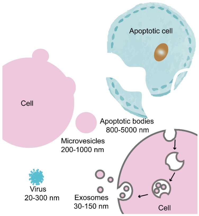
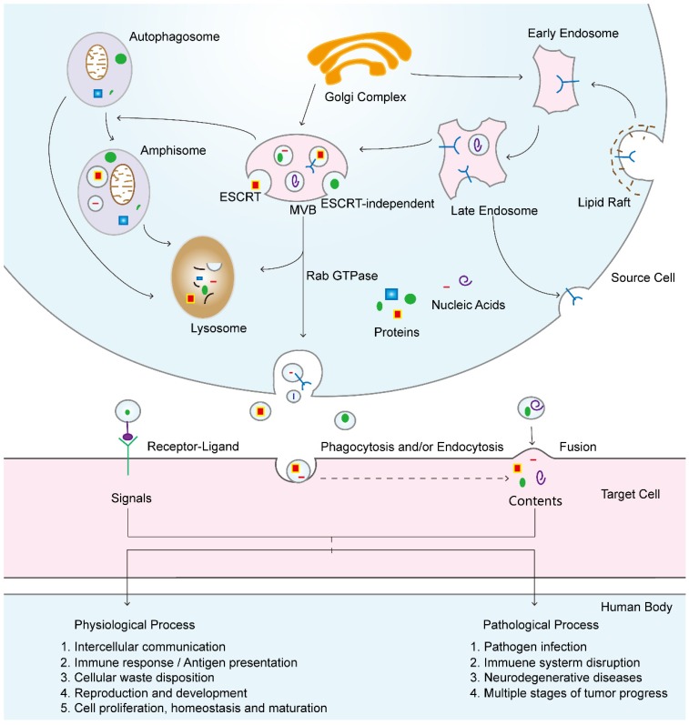
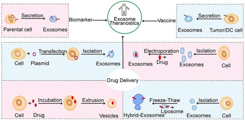
Exosomes are common membrane-bound nanovesicles that contain diverse biomolecules, such as lipids, proteins, and nucleic acids. Exosomes are derived from cells through exocytosis, are ingested by target cells, and can transfer biological signals between local or distant cells. Exosome secretion is a constitutive phenomenon that is involved in both physiological and pathological processes and determines both the exosomal surface molecules and the contents. Hence, we can exploit exosomes as biomarkers, vaccines and drug carriers and modify them rationally for therapeutic interventions. However, it is still a challenge to identify, isolate and quantify exosomes accurately, efficiently and selectively. Further studies on exosomes will explore their potential in translational medicine and provide new avenues for the creation of effective clinical diagnostics and therapeutic strategies; the use of exosomes in these applications can be called exosome theranostics. This review describes the fundamental processes of exosome formation and uptake. In addition, the physiological and pathological roles of exosomes in biology are also illustrated with a focus on how exosomes can be exploited or engineered as powerful tools in translational medicine.

Keywords: biomarker; drug delivery.; exosome; extracellular vesicle; translational medicine.


Conflict of interest statement

Competing Interests: The authors have declared that no competing interest exists.

Figures

Figure 1
Figure 1
Schematic representation of the biogenesis of apoptotic bodies, microvesicles and exosomes. Apoptotic bodies are vesicles that separate from post-apoptotic cells. Microvesicles are derived from the plasma membranes of most cell types. Exosomes are EVs of endocytic origin that are derived from most cell types. Viruses and exosomes are strikingly similar in structure and size.
Figure 2
Figure 2
Characterization of exosome-like vesicles. (A) Transmission electron micrograph of exosomes isolated from urine; scale bar, 400 nm. (B) Cryoelectron microscopy image showing extracellular vesicles secreted by MLP-29 cells; scale bar, 100 nm. (Reproduced with permission from reference . Copyright © 2008 American Chemical Society.) (C) Example of triple or higher-multiple vesicles; scale bar, 150 nm. (D) Percentage of each morphological category among the total number of vesicles. (E) Size distribution for each vesicle category. (C, D, E: reproduced with permission from reference . Copyright © 2017 Taylor & Francis Group.) (F) Electron micrograph of double membrane-bound exosomes in multivesicular bodies (MVBs); inward invagination (arrows) in the MVB membrane indicates the beginning of exosome biogenesis, scale bar, 100 nm. (Reproduced from reference . Copyright © 2011 American Heart Association, Inc.)
Figure 3
Figure 3
Exosomal biogenesis and internalization mechanisms and their roles in physiological and pathological processes. Exosomes are formed by inward budding from the endosomal membrane, which leads to the formation of multivesicular bodies (MVBs). MVBs can be fated for lysosomal degradation or fusion with the plasma membrane, which is associated with the release of exosomes. In addition, MVBs also participate in autophagosome maturation as endocytic fusion partners that meet with autophagosomes. Target cells internalize exosomes by three methods, which can facilitate the signaling and content delivery from source to target cells, thus mediating the progression of many physiological and pathological processes.
Figure 4
Figure 4
Schematic representation of the primary strategies of exosome modification and therapeutic intervention in translational medicine. Exosomes have the potential to serve as biomarkers and vaccine and drug carriers engineered by different methods. Such methods include the transfer of target peptide-expressing plasmids into cells to generate exosomes with a target ligand, the direct loading of drugs into exosomes by electroporation, the extrusion of drug-loaded cells through a series of filters with diminishing pore sizes to generate exosome-mimetic vesicles, and the fusion of exosomes with modified liposomes via the freeze-thaw method to create hybrid exosomes.
Figure 5
Figure 5
Exosome theranostics in pancreatic cancer. (A, B, C) Exosomes derived from the serum of pancreatic cancer (PC) patients initiate premetastatic niche formation in the liver, as evaluated by liver weight and the percentage of mCherry+ PAN02 cells in mice. (Reproduced with permission from reference . Copyright © 2015 Nature Publishing Group.) (D, E) Exosomes with surface-anchored glypican 1 (GPC1) proteins from the serum of PC patients are used as biomarkers to detect early PC. (Reproduced with permission from reference . Copyright © 2015 Nature Publishing Group.) (F) Change in the exosomal miRNAs level of the normal controls and PC patients. (Reproduced with permission from reference . Copyright © 2017 Elsevier) (G) HSP70/Bag-4+ exosomes that specifically induced the migration of natural killer cells. (Reproduced from reference . Copyright © 2005 American Association for Cancer Research.) (H, I, J) Exosomes that are loaded with short interfering RNA restrain PANC-1 tumor growth. (Reproduced with permission from reference . Copyright © 2017 Nature Publishing Group.)
Figure 6
Figure 6
Tumor metastases formation mediated by exosomes and metastatic tumor cell capturing in situ by a scaffold embedded with EVs (M-Trap) in an ovarian cancer dissemination model. (A) Schematic representation of tumor metastases. (B) Schematic of the peritoneal anatomy of mice, indicating M-Trap implantation. (C) Representative bioluminescent distribution of metastatic cells in the natural pattern of dissemination (D), in the presence of an empty scaffold (E) and in the presence of M-Trap. (B, C, D, E were reproduced from reference . Copyright © 2015 Oxford University Press.)

References

    1. Ludwig A-K, Giebel B. Exosomes: Small vesicles participating in intercellular communication. Int J Biochem Cell Biol. 2012;44:11–5. - PubMed
    1. Kaushik S, Cuervo AM. Proteostasis and aging. Nat Med. 2015;21:1406–15. - PubMed
    1. Christianson HC, Svensson KJ, van Kuppevelt TH, Li J-P, Belting M. Cancer cell exosomes depend on cell-surface heparan sulfate proteoglycans for their internalization and functional activity. Proc Natl Acad Sci. 2013;110:17380–5. - PMC - PubMed
    1. Frühbeis C, Fröhlich D, Kuo WP, Amphornrat J, Thilemann S, Saab AS. et al. Neurotransmitter-Triggered Transfer of Exosomes Mediates Oligodendrocyte-Neuron Communication. PLoS Biol. 2013;11:e1001604. - PMC - PubMed
    1. Raposo G, Nijman HW, Stoorvogel W, Liejendekker R, Harding CV, Melief CJ. et al. B lymphocytes secrete antigen-presenting vesicles. J Exp Med. 1996;183:1161–72. - PMC - PubMed

Publication types