

In Vitro Detection of Caries Around Amalgam Restorations Using Four Different Modalities
- PMID: 29290839
- PMCID: PMC5738745
- DOI: 10.2174/1874210601711010609
In Vitro Detection of Caries Around Amalgam Restorations Using Four Different Modalities
Abstract
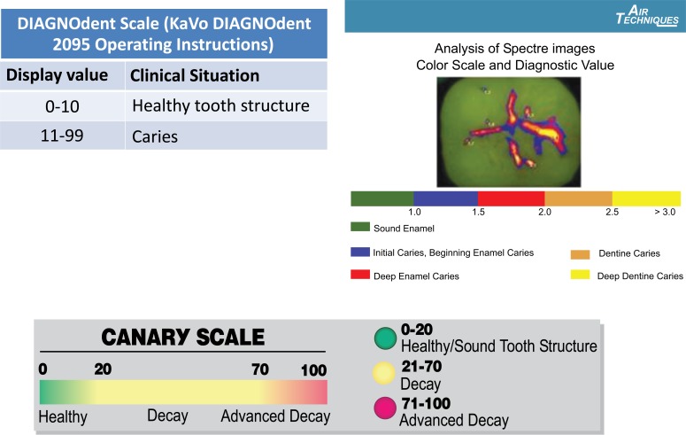
Objective: The aim of this study was to evaluate the ability of PTR-LUM (The Canary System, CS), laser fluorescence (DIAGNOdent, DD), LED fluorescence (Spectra), and visual inspection (ICDAS II) to detect natural decay around bonded amalgam restorations in vitro.
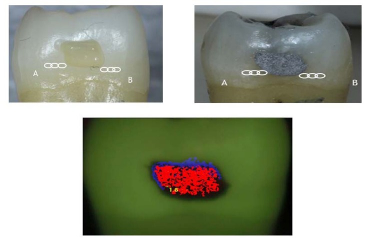
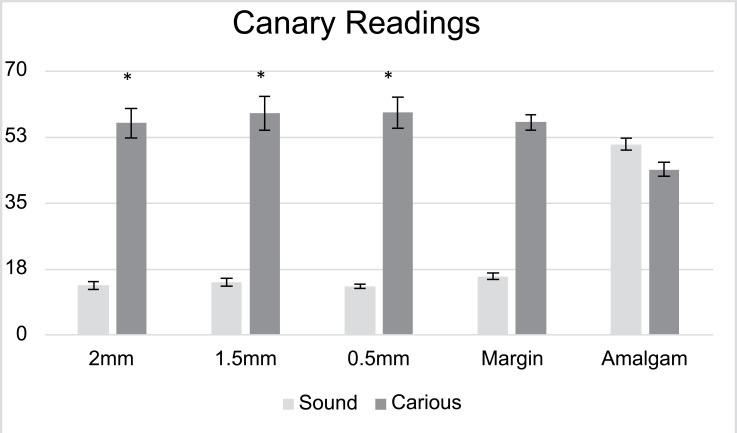
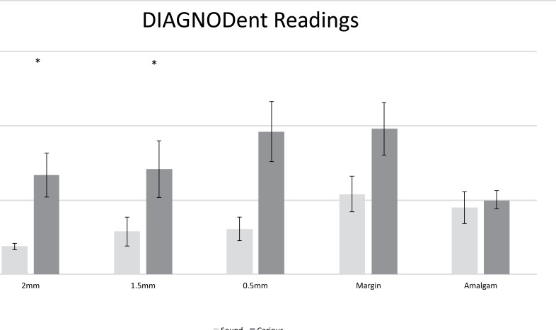
Methods: Seventeen extracted human molars and premolars, consisting of visually healthy (n=5) and natural cavitated (n=12) teeth were selected. For the carious teeth, caries was removed leaving some decayed tissue on the floor and or wall of the preparation. For sound teeth, 3 mm. deep cavity preparations were made and teeth were restored with bonded-amalgam restorations. Thirty-six sites (13 sound sites; 23 carious sites) were selected. CS and DD scans were performed in triplicate at 2, 1.5, 0.5, and 0 mm away from the margin of the restoration (MOR). Spectra images were captured for the entire surface, and dentists blinded to the samples provided ICDAS II scoring.
Results: Canary Numbers (Mean±SE) for healthy and carious sites at 2, 1.5, 0.5, and 0 mm from the MOR ranged from 12.9±0.9 to 15.4±0.9 and 56.1±4.0 to 56.3±2.0, respectively. DD peak values for healthy and carious sites ranged from 4.7±0.5 to 13.5±2.99, and 16.7±3.7 to 24.5±4.4, respectively. For CS and DD, sensitivity/specificity for sites at 2.0, 1.5, 0.5, 0 mm ranged from 0.95-1.0/0.85-1.0, and 0.45-0.74/0.54-1.0, respectively. For ICDAS II, sensitivity and specificity were 1.0 and 0.17, respectively. For Spectra, data and images were inconclusive due to signal intereference from the amalgam restoration.
Conclusions: Using this in-vitro model, CS and DD were able to differentiate between sound and carious tissue at the MOR, but larger variation, less reliability, and poorer accuracy was observed for DD. Therefore, CS has the potential to detect secondary caries around amalgam restorations more accurately than the other investigated modalities.
Keywords: Amalgam restorations; Canary System (CS); LED fluorescence (Spectra); Laser fluorescence; Margin of the restoration (MOR); Visual inspection (ICDAS II).
Figures

References
-
- Kidd E.A., Toffenetti F., Mjör I.A. Secondary caries. Int. Dent. J. 1992;42(3):127–138. - PubMed
LinkOut - more resources
Full Text Sources
Other Literature Sources
Miscellaneous