Distal Humeral Fractures-Current Concepts
- PMID: 29290875
- PMCID: PMC5721312
- DOI: 10.2174/1874325001711011353
Distal Humeral Fractures-Current Concepts
Abstract
Background: Distal humerus fractures constitute 2% of all fractures in the adult population. Although historically, these injuries have been treated non-operatively, advances in implant design and surgical technique have led to improved outcomes following operative fixation.
Methods: A literature search was performed and the authors' personal experiences are reported.
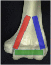
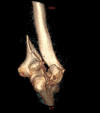
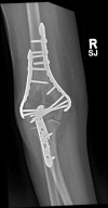
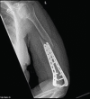
Results: This review has discussed the anatomy, classifications, treatment options and surgical techniques in relation to the management of distal humeral fractures. In addition, we have discussed controversial areas including the choice of surgical approach, plate orientation, transposition of the ulnar nerve and the role of elbow arthroplasty.
Conclusion: Distal humeral fractures are complex injuries that require a careful planned approach, when considering surgical fixation, to restore anatomy and achieve good functional outcomes.
Keywords: Anatomy; Distal humerus fracture; Elbow; Fracture fixation; Open reduction internal fixation; Total elbow arthroplasty.
Figures

References
-
- Frankle M.A., Herscovici D., Jr, DiPasquale T.G., Vasey M.B., Sanders R.W. A comparison of open reduction and internal fixation and primary total elbow arthroplasty in the treatment of intraarticular distal humerus fractures in women older than age 65. J. Orthop. Trauma. 2003;17(7):473–480. doi: 10.1097/00005131-200308000-00001. - DOI - PubMed
-
- McKee M.D., Jupiter J.B. Fractures of the distal humerus. 3 rd ed. Philadelphia: Lippincott: Skeletal trauma.; 2002. pp. 765–782.
Publication types
LinkOut - more resources
Full Text Sources
Other Literature Sources
