Embryonal rhabdomyosarcoma in the maxillary sinus with orbital involvement in a pediatric patient: Case report
- PMID: 29291204
- PMCID: PMC5740190
- DOI: 10.12998/wjcc.v5.i12.440
Embryonal rhabdomyosarcoma in the maxillary sinus with orbital involvement in a pediatric patient: Case report
Abstract
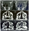
This report presents a case of embryonal rhabdomyosarcoma (eRMS) located in the left maxillary sinus and invading the orbital cavity in a ten-year-old male patient who was treated at a referral hospital. The images provided from the computed tomography showed a heterogeneous mass with soft-tissue density, occupying part of the left half of the face inside the maxillary sinus, and infiltrating and destroying the bone structure of the maxillary sinus, left orbit, ethmoidal cells, nasal cavity, and sphenoid sinus. An analysis of the histological sections revealed an undifferentiated malignant neoplasm infiltrating the skeletal muscle tissue. The immunohistochemical analysis was positive for the antigens: MyoD1, myogenin, desmin, and Ki67 (100% positivity in neoplastic cells), allowing the identification of the tumour as an eRMS. The treatment protocol included initial chemotherapy followed by radiotherapy and finally surgery. The total time of the treatment was nine months, and in 18-mo of follow-up period did not show no local recurrences and a lack of visual impairment.
Keywords: Chemotherapy; Embryonal rhabdomyosarcoma; Maxillary sinus; Oncology; Pediatrics.
Conflict of interest statement
Conflict-of-interest statement: We, the authors of this paper “Embryonal rhabdomyosarcoma in the maxillary sinus with orbital involvement in a pediatric patient: Case report”, stating that we participate sufficiently in the design of the study and development of this work and we take public responsibility on it and we delegate to the World Journal of Clinical Cases the copyright upon acceptance of the publication of this. The authors undersigned declare no conflict of interest regarding this manuscript, as well as the information it contains.
Figures


References
-
- Reilly BK, Kim A, Peña MT, Dong TA, Rossi C, Murnick JG, Choi SS. Rhabdomyosarcoma of the head and neck in children: review and update. Int J Pediatr Otorhinolaryngol. 2015;79:1477–1483. - PubMed
-
- Garay M, Chernicoff M, Moreno S, Pizzi de Parra N, Oliv, J, Apréa G. Rabdomiosarcoma alveolare congenito in un neonato. Eur J Pediatr Dermatol. 2004;14:9–12.
Publication types
LinkOut - more resources
Full Text Sources
Other Literature Sources

