. 2018 Mar;1860(3):792-800.
doi: 10.1016/j.bbamem.2017.12.019. Epub 2017 Dec 30.

Binding, folding and insertion of a β-hairpin peptide at a lipid bilayer surface: Influence of electrostatics and lipid tail packing

Affiliations

Binding, folding and insertion of a β-hairpin peptide at a lipid bilayer surface: Influence of electrostatics and lipid tail packing

Keon A Reid et al. Biochim Biophys Acta Biomembr. 2018 Mar.

Abstract

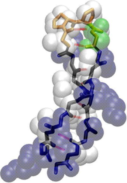
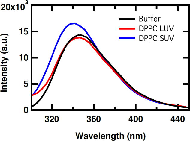
Antimicrobial peptides (AMPs) act as host defenses against microbial pathogens. Here we investigate the interactions of SVS-1 (KVKVKVKVdPlPTKVKVKVK), an engineered AMP and anti-cancer β-hairpin peptide, with lipid bilayers using spectroscopic studies and atomistic molecular dynamics simulations. In agreement with literature reports, simulation and experiment show preferential binding of SVS-1 peptides to anionic over neutral bilayers. Fluorescence and circular dichroism studies of a Trp-substituted SVS-1 analogue indicate, however, that it will bind to a zwitterionic DPPC bilayer under high-curvature conditions and folds into a hairpin. In bilayers formed from a 1:1 mixture of DPPC and anionic DPPG lipids, curvature and lipid fluidity are also observed to promote deeper insertion of the fluorescent peptide. Simulations using the CHARMM C36m force field offer complementary insight into timescales and mechanisms of folding and insertion. SVS-1 simulated at an anionic mixed POPC/POPG bilayer folded into a hairpin over a microsecond, the final stage in folding coinciding with the establishment of contact between the peptide's valine sidechains and the lipid tails through a "flip and dip" mechanism. Partial, transient folding and superficial bilayer contact are seen in simulation of the peptide at a zwitterionic POPC bilayer. Only when external surface tension is applied does the peptide establish lasting contact with the POPC bilayer. Our findings reveal the influence of disruption to lipid headgroup packing (via curvature or surface tension) on the pathway of binding and insertion, highlighting the collaborative effort of electrostatic and hydrophobic interactions on interaction of SVS-1 with lipid bilayers.

Keywords: Curvature; Fluorescence spectroscopy; Lipid bilayers; Liposome; SVS-1; Surface tension.


Figures

Figure 1
Figure 1
Model of folded SVS-1 structure created using Visual Molecular Dynamics (VMD) [29]. Peptide backbone (black) is represented as a licorice, and VDPLPT turn motif colored in orange. Peptide sidechains are presented as VDW beads – hydrophobic valines and prolines (white), basic lysine (blue) and polar threonine (green). The intermolecular backbone hydrogen bonds are drawn in red.
Figure 2
Figure 2
Far-UV CD spectra of 50 μM SVS-1 mixed with 1:1 DPPC/DPPG LUV at molar ratios between 1:100 and 1:0.5 in 10 mM sodium phosphate buffer, 150 mM NaCl pH 7.4 at 20 °C.
Figure 3
Figure 3
Far-UV CD spectra of 60 μM SVS-V8W in 10 mM sodium phosphate buffer and 150 mM NaCl pH 7.4 (black), 2.5 mM DPPC LUV (red), and 2.5 mM DPPC SUV (blue) at 20 °C.
Figure 4
Figure 4
Fluorescence spectra of 60 μM SVS-V8W in 10 mM sodium phosphate buffer and 150 mM NaCl pH 7.4 (black), 2.5 mM DPPC LUV (red), and 2.5 mM DPPC SUV (blue) at 20 °C excited at 280 nm.
Figure 5
Figure 5
Fluorescence spectra of 60 μM SVS-V8W in 10 mM sodium phosphate buffer and 150 mM NaCl pH 7.4 (black), 2.5 mM 1:1 DPPC/DPPG LUV (red), and 2.5 mM 1:1 DPPC/DPPG SUV (blue) at 20 °C excited at 280 nm.
Figure 6
Figure 6
Normalized fluorescence spectra of 60 μM SVS-V8W in 10 mM sodium phosphate buffer and 150 mM NaCl pH 7.4 (black), 2.5 mM 1:1 DPPC/DPPG SUV (blue dash) and 2.5 mM 1:1 DPPC/DPPG LUV (red) at 20 °C and 2.5 mM 1:1 DPPC/DPPG LUV (red dash) at 65 °C excited at 280 nm. Inset: Intensity at the wavelength maximum, 348 nm, of 60 μM SVS-V8W in 2.5 mM 1:1 DPPC/DPPG LUV acquired every 5 °C from 5 to 65 °C during the course of a temperature-dependent experiment in a 1 cm pathlength cell. The continuous line represents the best fit of the data to a sigmoid (Equation 1).
Figure 7
Figure 7
Number of native hairpin SVS-1 hydrogen bonds in 1000 ns solution-phase simulation, with corresponding peptide snapshots. Folded peptides are drawn in New Cartoon method using VMD. The plot has been smoothed, showing averages over 1 ns.
Figure 8
Figure 8
Bilayer contact fluctuations and hydrogen bond formation (folding dynamics) of SVS-1 in presence of a zwitterionic POPC bilayer (500 ns) and an anionic POPC/POPG bilayer (1500 ns). Fluctuations track distances of terminal sites and center of turn to the closest acyl tail carbon. The atoms are colored based on description in Methods (See cartoon in upper right panel). Hydrogen bond order from 1-8 (starting from turn to the tail-end) are represented in order by the colors: black, red, green, blue, yellow, brown, purple and magenta (See cartoon in lower right panel). The curves represent averages smoothed over 1 ns.
Figure 9
Figure 9
Snapshots from the 1500 ns simulation of SVS-1 interaction with 1:1 POPC/POPG bilayer. Figure A shows partially folded structure with strong interactions of lysine sidechains with headgroups at 1040 ns. Figure B highlights the initiation of the “flip and dip” mechanism, where the turn buries beneath the headgroups (at 1075 ns). Figure C depicts endpoint of the “flip and dip” mechanism after 50 ns (1130 ns). Figure D (top-down view) represents SVS-1 with valines inserted and lysines facing upwards into solution (end of 1500 ns simulation). Red, green, and black sites as defined in Figure 8 are shown as spheres. SVS-1 sidechains are depicted as a licorice: valine(blue), lysine(green), proline(orange) and threonine(yellow). Lipids are represented as dotted in VMD drawing method. Some lipids are removed for Figures A–C for clarity of mechanism. Water and ions removed for clarity.
Figure 10
Figure 10
Number of native hydrogen bonds formed (left) and number of valine-to-tail contacts (right) for a folded or unfolded SVS-1 peptide in the presence of an anionic or zwitterionic bilayer (at 323 K) using C36m, as defined in section 2.2.4. Simulations for each of the starting configurations, folded (f) or unfolded (uf), were performed under applied surface tension factors of 10 or 30 mN/m, respectively. Curves represent averages smoothed over 1 ns.

References

    1. Yeaman MR, Yount NY. Mechanisms of antimicrobial peptide action and resistance. Pharmacological Reviews. 2003;55(1):27–55. - PubMed
    1. Bond PJ, Khalid S. Antimicrobial and Cell-Penetrating Peptides: Structure, Assembly and Mechanisms of Membrane Lysis via Atomistic and Coarse-Grained Molecular Dynamic Simulations. Protein and Peptide Letters. 2010;17(11):1313–1327. - PubMed
    1. Nakatsuji T, Gallo RL. Antimicrobial Peptides: Old Molecules with New Ideas. J Invest Dermatol. 2012;132(3):887–898. - PMC - PubMed
    1. Giuliani A, Pirri G, Nicoletto S. Antimicrobial peptides: an overview of a promising class of therapeutics. Cent Eur J Biol. 2007;2(1):1–33.
    1. Sinthuvanich C, Veiga AS, Gupta K, Gaspar D, Blumenthal R, Schneider JP. Anticancer β-Hairpin Peptides: Membrane-Induced Folding Triggers Activity. JACS. 2012;134(14):6210–6217. - PMC - PubMed

Publication types

LinkOut - more resources