. 2018 Feb:115:94-103.
doi: 10.1016/j.yjmcc.2017.12.014. Epub 2017 Dec 30.

Serotonin receptor 2B signaling with interstitial cell activation and leaflet remodeling in degenerative mitral regurgitation

Affiliations

Serotonin receptor 2B signaling with interstitial cell activation and leaflet remodeling in degenerative mitral regurgitation

Kathryn H Driesbaugh et al. J Mol Cell Cardiol. 2018 Feb.

Abstract

Aims: Mitral valve interstitial cells (MVIC) play an important role in the pathogenesis of degenerative mitral regurgitation (MR) due to mitral valve prolapse (MVP). Numerous clinical studies have observed serotonin (5HT) dysregulation in cardiac valvulopathies; however, the impact of 5HT-mediated signaling on MVIC activation and leaflet remodeling in MVP have been investigated to a limited extent. Here we test the hypothesis that 5HT receptors (5HTRs) signaling contributes to MVP pathophysiology.

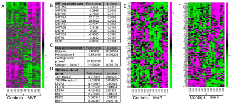
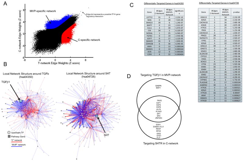
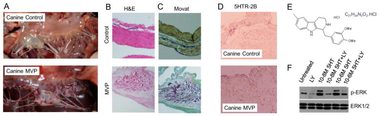
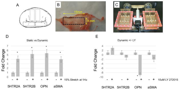
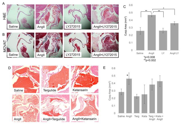
Methods and results: Diseased human MV leaflets were obtained during cardiac surgery for MVP; normal MV leaflets were obtained from heart transplants. MV RNA was used for microarray analysis of MVP patients versus control, highlighting genes that indicate the involvement of 5HTR pathways and extracellular matrix remodeling in MVP. Human MV leaflets were also studied in vitro and ex vivo with biomechanical testing to assess remodeling in the presence of a 5HTR2B antagonist (LY272015). MVP leaflets from Cavalier King Charles Spaniels were used as a naturally acquired in vivo model of MVP. These canine MVP leaflets (N=5/group) showed 5HTR2B upregulation. This study also utilized CB57.1ML/6 mice in order to determine the effect of Angiotensin II infusion on MV remodeling. Histological analysis showed that MV thickening due to chronic Angiotensin II remodeling is mitigated by a 5HTR2B antagonist (LY272015) but not by 5HTR2A inhibitors.

Conclusion: In humans, MVP is associated with an upregulation in 5HTR2B expression and increased 5HT receptor signaling in the leaflets. Antagonism of 5HTR2B mitigates MVIC activation in vitro and MV remodeling in vivo. These observations support the view that 5HTR signaling is involved not only in previously reported 5HT-related valvulopathies, but it is also involved in the pathological remodeling of MVP.

Keywords: Angiotensin; Cardiovascular disease; Mitral valve; Physiology; Serotonin; Surgery.


Figures

Figure 1
Figure 1. Human microarray analysis and identification of 5HT signaling pathways in MVP patients
(A) Heat maps showing the relative expression levels of 1883 differentially-expressed transcripts from the MVP and control samples (N=4). Ordering of the genes and samples is based on a hierarchical clustering and the expression values across rows are Z-score normalized for visualization purposes only. Green indicates a positive fold change while purple indicates a negative fold change. (B) (C) and (D) Tables showing selected genes differentially expressed between MVP and Controls for 5HT, ECM, and TGFβ signaling, respectively. Fold changes and p value are indicated. (E) and (F) Heat maps showing the relative expression levels of genes from the MVP and control expression samples for the TGFB signaling pathway (hsa04350) or the serotonin pathways (hsa04726).
Figure 2
Figure 2. Regulatory Network reconstruction in MVP vs. Controls
(A) A plot of the edge weights predicted by PANDA when reconstructing a regulatory network using the MVP or control expression data samples. On the plot, each point represents a potential transcription factor to target gene regulatory relationship. Regulatory edges that were identified as specific to either the MVP-network or the C-network are shown in red and blue, respectively. (B) Visualization of the MVP or C-specific sub-networks in which a member of either the TGFβ signaling pathway (hsa04350, left panel) or the serotonin pathway (hsa04726, right panel) are target genes. (C) The number of edges targeting members of the TGFβ signaling pathway or the serotonin signaling pathway in each of the two identified MVP-specific and C-specific sub-networks. The significance of any differential-targeting of these genes comparing the sub-networks is also shown. (D) A Venn diagram of the transcription factor regulators identified as targeting 5HT in the MVP-network and TGFβ in the C-network.
Figure 3
Figure 3. MVP is associated with increased protein expression of 5HTR2B
Representative immunohistochemistry staining of 4 μm thick cross sections of human MV leaflets surgically resected from patients with MR due to MVP and controls (N=4 group) using (A) H&E, (B) MOVAT staining. (C) Quantitative PCR analysis for 5HTRA and 5HTR2B expression in N=5 independent control and MVP MV specimens. *p=0.05. IHC with (D) anti-5HTR2A (E) anti-5HTR2B, (F) anti-αSMA, and (G) anti-OPN antibodies, respectively. Magnification, 63X.
Figure 4
Figure 4. Canine MV disease shows a marked increase in 5HTR2B expression
(A) Representative intra-op images of normal canine MV and canine valve with MVP presentations. Representative (B) H&E staining and (C) Modified Movat Pentachrome staining of control and MVP canine tissue (N=4) (Magnification 40x). (D) Immunohistochemistry staining of 4 μm thick cross sections of canine MV leaflets surgically resected from MVP and controls (N=4 group) using an anti-5HTR2B antibody. Magnification 40X. (E) LY 272015 and (F) Isolated canine MVICs were serum -starved overnight and pre-treated for 30 minutes with vehicle or 100 nM in serum free Advanced DMEM media, followed by treatment with the indicated dose of 5HT for 5 minutes. Protein lysates were analyzed by immunoblot analysis using anti-phospho-ERK and anti-ERK antibodies, respectively.
Figure 5
Figure 5. Ex vivo mechanical stimulation induces 5HTR expression
(A–C) Processing of human MV tissue and the uniaxial bioreactor (D) Control MV tissues were loaded into the BioReactor and maintained in static or dynamic (1Hz, 15% stretch) conditions for 6 days. RNA was isolated from the tissue, reverse transcribed, and qPCR analysis was performed using primers specific for 5HTR2A, 5HTR2B, OPN and αSMA. Control MV tissues were exposed to (E) 10 μM 5HT +/− 100 μM LY 272015 for 6 days under controlled biomechanical stimulation (1Hz, 15% stretch). RNA was isolated from the tissue, reverse transcribed, and qPCR analysis was performed using primers specific for 5HTR2A, 5HTR2B, OPN, and αSMA, *p=0.05, **p=0.01.
Figure 6
Figure 6. Angiotensin II infusion promotes remodeling of the mitral valve tissue in mice which can be partially blocked by 5HTR2B inhibition
(A) Representative H&E and (B) Modified Movat Pentachrome staining of cross sections of MV from mice hearts harvested 28 days after saline or Ang II chronic infusion with or without treatment of 10 μM LY 272015 (Magnification 20x). (C) Quantitative analysis of the average MV leaflet section area for each treatment group +/− SE. *p<0.008; **p<0.002. (D) Representative H&E of MV from mice hearts harvested 28 days after saline or Ang II chronic infusion with or without treatment Terguride or Ketansarin. (E) Quantitative analysis of the average MV leaflet section area for each treatment group indicated in D +/− SE. *p<0.002.

References

    1. Sainger R, Grau JB, Branchetti E, Poggio P, Seefried WF, Field BC, et al. Human myxomatous mitral valve prolapse: role of bone morphogenetic protein 4 in valvular interstitial cell activation. J Cell Physiol. 2012;227:2595–2604. doi: 10.1002/jcp.22999. - DOI - PMC - PubMed
    1. Guy TS, Hill AC. Mitral valve prolapse. Annu Rev Med. 2012;63:277–292. doi: 10.1146/annurev-med-022811-091602. - DOI - PubMed
    1. Freed LA, Levy D, Levine RA, Evans JC, Larson MG, Fuller DL, et al. Mitral valve prolapse and atrial septal aneurysm: an evaluation in the Framingham Heart Study. Am J Cardiol. 2002;89:1326–1329. - PubMed
    1. Nishimura RA, Otto CM, Bonow RO, Carabello BA, Erwin JP, Guyton RA, et al. 2014 AHA/ACC guideline for the management of patients with valvular heart disease: executive summary: a report of the American College of Cardiology/American Heart Association Task Force on Practice Guidelines. J Am Coll Cardiol. 2014;63:2438–2488. doi: 10.1016/j.jacc.2014.02.537. - DOI - PubMed
    1. Salhiyyah K, Yacoub MH, Chester AH. Cellular mechanisms in mitral valve disease. J Cardiovasc Transl Res. 2011;4:702–709. doi: 10.1007/s12265-011-9318-7. - DOI - PubMed

Publication types

MeSH terms