Review
. 2018 Aug:330:142-150.
doi: 10.1016/j.cellimm.2017.12.009. Epub 2017 Dec 26.

Muscularis macrophages: Key players in intestinal homeostasis and disease

Affiliations
Review

Muscularis macrophages: Key players in intestinal homeostasis and disease

Sebastiaan De Schepper et al. Cell Immunol. 2018 Aug.

Abstract

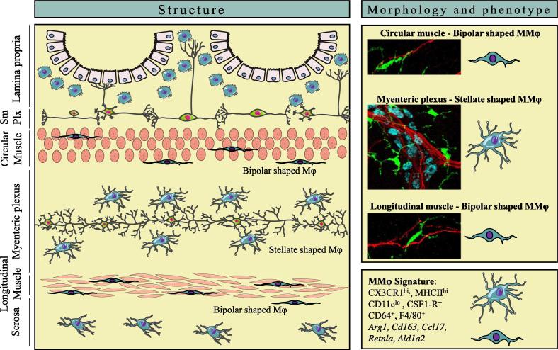
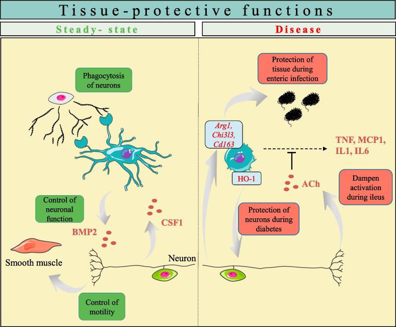
Macrophages residing in the muscularis externa of the gastrointestinal tract are highly specialized cells that are essential for tissue homeostasis during steady-state conditions as well as during disease. They are characterized by their unique protective functional phenotype that is undoubtedly a consequence of the reciprocal interaction with their environment, including the enteric nervous system. This muscularis macrophage-neuron interaction dictates intestinal motility and promotes tissue-protection during injury and infection, but can also contribute to tissue damage in gastrointestinal disorders such as post-operative ileus and gastroparesis. Although the importance of muscularis macrophages is clearly recognized, different aspects of these cells remain largely unexplored such their origin, longevity and instructive signals that determine their function and phenotype. In this review, we will discuss the phenotype, functions and origin of muscularis macrophages during steady-state and disease conditions. We will highlight the bidirectional crosstalk with neurons and potential therapeutic strategies that target and manipulate muscularis macrophages to restore their protective signature as a treatment for disease.

Keywords: Enteric nervous system; Gastrointestinal disorders; Intestinal macrophage; Intestinal muscularis externa; Neuro-immune interactions; Tissue-macrophage ontogeny.


Figures

Fig. 1
Fig. 1
The localization and phenotype of muscularis macrophages. (Left panel) Anatomical overview of macrophage (Mφ) distribution in different layers of the gastrointestinal tract. (Upper right panel) Mφ (green, CX3CR1) located in the muscularis externa (MMφ) have a distinct morphology dependent on their position within circular and longitudinal muscle layers or myenteric plexus. MMφ in the myenteric plexus resemble microglia with a ramified, stellate shaped morphology and closely contact enteric neurons (red, TUBB3) and myenteric ganglia (blue, HUC/D). (Lower right panel) MMφ express typical Mφ-specific surface markers such as CX3CR1, CD64 and F4/80. They are characterized by their tissue-protective genetic signature, such as the expression of Arg1, Cd163, Ccl17, Retnla and Ald1a2. SmPlx = submucosal plexus. (For interpretation of the references to color in this figure legend, the reader is referred to the web version of this article.)
Fig. 2
Fig. 2
Muscularis macrophages contribute to tissue-protective functions during steady-state and disease. (Left) Muscularis macrophages (MMφ) are actively communicating with surrounding neurons, being imprinted by neuronal-derived CSF-1 for their maintenance and development. Conversely, MMφ sustain neuronal function and motility via BMP2 secretion. In addition, MMφ participate in the phagocytosis of surrounding neurons. (Right) MMφ are involved in tissue protection during disease conditions. During postoperative ileus, vagus nerve stimulation can activate enteric neurons, leading to increased production of acetylcholine, and dampening of MMφ. This reduces inflammation and accelerates the recovery of motility. Moreover, MMφ exert a M2-like signature that is enhanced upon enteric infection and express the enzyme heme oxygenase-1 (HO-1), that exerts a protective effect on neurons during diabetes.

References

    1. Davies L.C., Jenkins S.J., Allen J.E., Taylor P.R. Tissue-resident macrophages. Nat. Immunol. 2013;14:986–995. - PMC - PubMed
    1. Hussell T., Bell T.J. Alveolar macrophages: plasticity in a tissue-specific context. Nat. Rev. Immunol. 2014;14:81–93. - PubMed
    1. Rivollier A., He J., Kole A., Valatas V., Kelsall B.L. Inflammation switches the differentiation program of Ly6Chi monocytes from antiinflammatory macrophages to inflammatory dendritic cells in the colon. J. Exp. Med. 2012;209:139–155. - PMC - PubMed
    1. Parkhurst C.N., Yang G., Ninan I., Savas J.N., Yates J.R., 3rd, Lafaille J.J., Hempstead B.L., Littman D.R., Gan W.B. Microglia promote learning-dependent synapse formation through brain-derived neurotrophic factor. Cell. 2013;155:1596–1609. - PMC - PubMed
    1. Gosselin D., Link V.M., Romanoski C.E., Fonseca G.J., Eichenfield D.Z., Spann N.J., Stender J.D., Chun H.B., Garner H., Geissmann F., Glass C.K. Environment drives selection and function of enhancers controlling tissue-specific macrophage identities. Cell. 2014;159:1327–1340. - PMC - PubMed

Publication types