. 2018 Feb 7;26(2):524-541.
doi: 10.1016/j.ymthe.2017.11.019. Epub 2017 Dec 5.

Triple Vectors Expand AAV Transfer Capacity in the Retina

Affiliations

Triple Vectors Expand AAV Transfer Capacity in the Retina

Andrea Maddalena et al. Mol Ther. .

Abstract

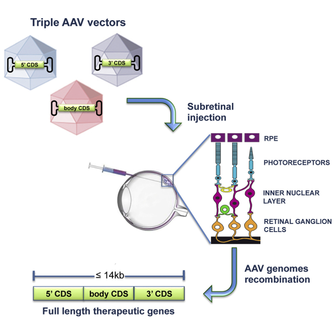
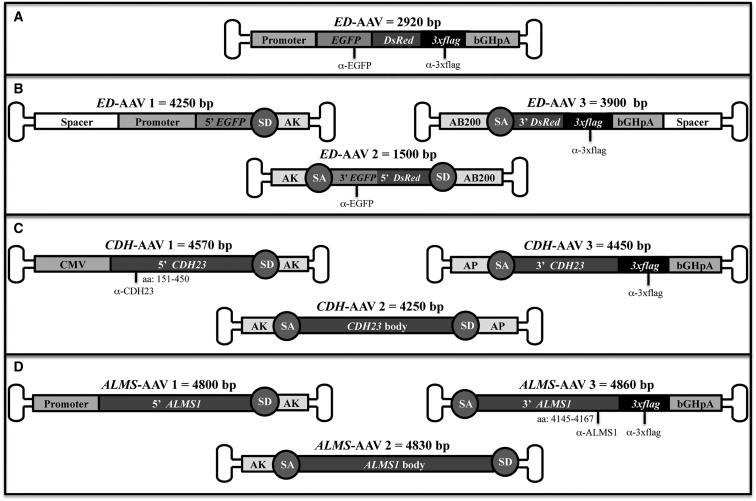
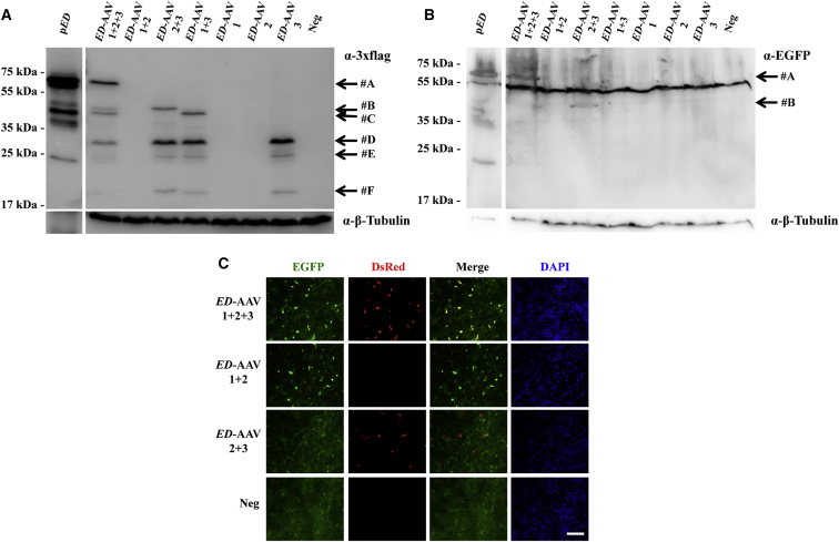
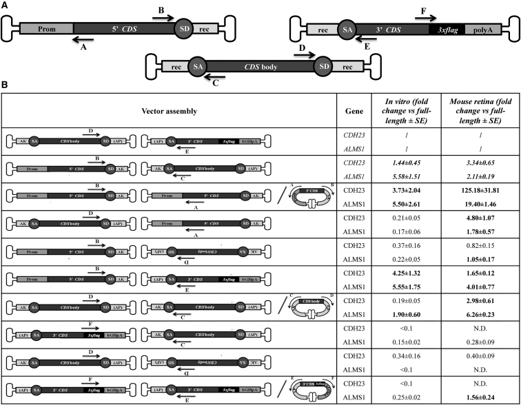
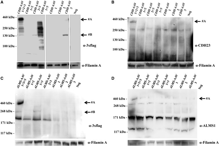
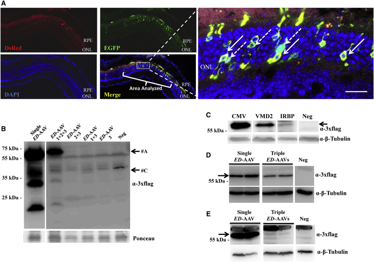
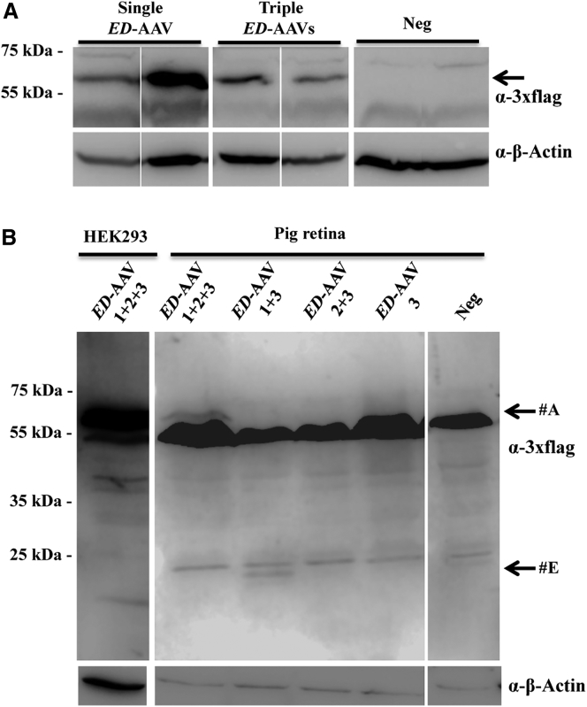
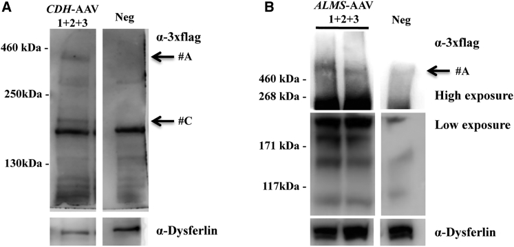
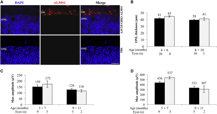
Retinal gene transfer with adeno-associated viral (AAV) vectors holds great promise for the treatment of inherited retinal degenerations (IRDs). One limit of AAV is its transfer capacity of about 5 kb, which can be expanded to about 9 kb, using dual AAV vectors. This strategy would still not suffice for treatment of IRDs such as Usher syndrome type 1D or Alström syndrome type I (ALMS) due to mutations in CDH23 or ALMS1, respectively. To overcome this limitation, we generated triple AAV vectors, with a maximal transfer capacity of about 14 kb. Transcriptomic analysis following triple AAV transduction showed the expected full-length products along a number of aberrant transcripts. However, only the full-length transcripts are efficiently translated in vivo. We additionally showed that approximately 4% of mouse photoreceptors are transduced by triple AAV vectors and showed correct localization of recombinant ALMS1. The low-photoreceptor transduction levels might justify the modest and transient improvement we observe in the retina of a mouse model of ALMS. However, the levels of transduction mediated by triple AAV vectors in pig retina reached 40% of those observed with single vectors, and this bodes well for further improving the efficiency of triple AAV vectors in the retina.

Keywords: AAV; gene therapy; large gene; retina; triple AAV.


Figures

None
Graphical abstract
Figure 1
Figure 1
Schematic Representation of Single and Triple AAV Vectors Single ED-AAV (A); triple ED-AAVs (B); triple CDH-AAVs (C); triple ALMS-AAVs (D). The position of the epitopes recognized by the antibodies used in this study is indicated. The vector genome size is depicted above each vector. bGHpA, bovine growth hormone polyadenylation signal; SD, splicing donor signal; SA, splicing acceptor signal; AP, alkaline phosphatase recombinogenic region; AK, F1 phage recombinogenic region; AB200, ABCA4 recombinogenic region; 3xflag, triple flag tag; CMV, cytomegalovirus; α-EGFP, anti-EGFP antibodies; α-3xflag, anti-3xflag antibodies; α-CDH23, anti-CDH23 antibodies; α-ALMS1, anti-ALMS1 antibodies.
Figure 2
Figure 2
Triple ED-AAV Vectors Efficiently Transduce ED In Vitro (A and B) Western blot (WB) analysis of lysates from HEK293 cells either transfected with pED or infected with triple AAV2/2 vectors encoding for ED and stained with α-3xflag (A) and α-EGFP (B) antibodies. The arrows on the right indicate the following protein products: #A, ED full-length protein; #B, from AAV 2 + 3; #C, from AAV 1 + 3; #F, from AAV 3; #D and #E, aggregates of AAV 3. α-3xflag, WB with anti-3xflag antibodies; α-EGFP, WB with anti-EGFP antibodies; α-β-Tubulin, WB with anti-β-Tubulin antibodies, used as loading control. Neg, not infected cells. The molecular weight ladder is depicted on the left; 20 and 200 μg of proteins for transfected and infected samples, respectively, were loaded. The WB images are representative of n = 4 independent experiments. (C) Fluorescent analysis of HEK293 cells infected with triple AAV2/2 vectors encoding for ED. The scale bar (200 μm) is depicted in the figure. DsRed, Discosoma red fluorescent protein; Merge: overlay of EGFP and DsRed images.
Figure 3
Figure 3
Transcriptomic Analysis following Transduction with Triple AAV Vectors (A) Schematic representation of triple CDH and ALMS-AAV vectors with primers used for real-time qPCR (A→F). Prom, promoter; CDS, coding sequence; polyA, polyadenylation signal; 3xflag, triple flag tag; SD, splicing donor signal; SA, splicing acceptor signal; rec, recombinogenic region. (B) Results of the real-time qPCR analysis performed on cDNAs from HEK293 cells or mouse eyecups treated with either CDH- or ALMS-AAV 1 + 2 + 3 vectors. The left column shows the vector assembly predicted on the basis of the set of primers used. The two columns on the right show the abundance of the PCR products relative to full-length detected by the D + E primers. Only products with a relative abundance >0.1 are shown. Highlighted in italic is the relative abundance of PCR products resulting from the desired AAV directional concatemerization. Highlighted in bold are the PCR products with a relative abundance >1.
Figure 4
Figure 4
Triple CDH- and ALMS-AAV Vectors Efficiently Transduce CDH23 and ALMS1 In Vitro (A and B) WB analysis of lysates from HEK293 cells infected with triple AAV2/2 vectors encoding for CDH23 and incubated with either anti-3xflag (A) or anti-CDH23 (B) antibodies. The arrows on the right indicate the following protein products: #A, full-length CDH23 protein; #B, from AAV 3. (C and D) WB analysis of lysates from HEK293 cells infected with triple AAV2/2 vectors encoding for ALMS1 and incubated with either anti-3xflag (C) or anti-ALMS1 (D) antibodies. The arrows on the right indicate the following protein products: #A, full-length ALMS1 protein; #B, from AAV 1 + 3. α-3xflag, WB with anti-3xflag antibodies; α-CDH23, WB with anti-CDH23 antibodies; α-ALMS1, WB with anti-ALMS1 antibodies; α-Filamin A, WB with anti-Filamin A antibodies, used as loading control. Neg, cells infected with control AAV2/2-CMV-EGFP vectors. The molecular weight ladder is depicted on the left, 100 (A), 250 (B), or 200 μg (C and D) of proteins were loaded.
Figure 5
Figure 5
Triple AAV Vectors Drive Full-Length Protein Expression in the Mouse Retina (A) Fluorescence analysis of retinal cryosections from C57BL/6J mice 2 months following subretinal injection of triple ED-AAV2/8 vectors under the control of the ubiquitous CMV promoter. The pictures with insets at higher magnification are representative of n = 5 injected eyes. Full arrows indicate EGFP and DsRed double-positive PRs. Dashed arrows indicate EGFP-only positive PRs. The scale bar (20 μm) is depicted in the figure. DsRed, Discosoma red fluorescent protein; Merge, overlay of EGFP, DsRed, and DAPI images; RPE, retinal pigmented epithelium; ONL, outer nuclear layer. (B) Western blot (WB) analysis of truncated products in eyecup lysates from C57BL/6J mice 2 months following subretinal injection of the combinations of ED-AAVs. The arrows indicate the following products: #A, ED full-length protein; #C, from AAV 1 + 3. (C) WB analysis of lysates from C57BL/6J eyecup 2 months following subretinal injection of triple AAV2/8 vectors encoding for ED under the control of the ubiquitous CMV, the RPE-specific VMD2, and the PR-specific IRBP promoters. The arrow indicates the full-length protein. (D) WB analysis of eyecup lysates from C57BL/6J mice 2 months following subretinal injection of single and triple AAV2/8 vectors encoding for ED under the control of the ubiquitous promoter CMV. (E) WB analysis of eyecup lysates from C57BL/6J mice 2 months following subretinal injection of single and triple AAV2/8 vectors encoding for ED under the control of the PR-specific promoter IRBP. CMV, cytomegalovirus; VMD2, vitelliform macular dystrophy 2; IRBP, human interphotoreceptors retinoid binding proteins. α-3xflag, WB with anti-3xflag antibodies; Ponceau, staining with Ponceau, used as loading control; α-β-Tubulin, WB with anti-β-Tubulin antibodies, used as loading control. Neg, lysates of eyecups following injection with PBS. The molecular weight ladder is depicted on the left; 100 (D), 150 (B and C), and 200 (E) μg of proteins were loaded.
Figure 6
Figure 6
Triple AAV Vectors Drive Full-Length Protein Expression in the Pig Retina (A) Western blot (WB) analysis of lysates from Large White pig retina 2 months following subretinal injection of either single or triple AAV2/8 vectors encoding for ED under the control of the PR-specific promoter IRBP. The arrow indicates the full-length protein. (B) WB analysis of truncated products in lysates from Large White pig retina 2 months following subretinal injection of the combinations of triple ED-AAVs. Lysates of HEK293 cells infected with an ED-AAV 1 + 2 + 3 are shown on the left as positive control. The arrows indicate the following products: #A, ED full-length protein; #E, probably from AAV 3. α-3xflag, WB with anti-3xflag antibodies; α-β-Actin, WB with anti-β-Actin antibodies, used as loading control. Neg, lysates of retinas following injection with PBS. The molecular weight ladder is depicted on the left; 200 μg of proteins were loaded.
Figure 7
Figure 7
Subretinal Administration of Triple AAV Vectors Results in Full-Length CDH23 and ALMS1 Protein Expression in the Mouse Retina (A) Western blot (WB) analysis of eyecup lysates from C57BL/6J mice 3 months following subretinal injection of triple AAV2/8 encoding for CDH23 under the control of the ubiquitous CMV promoter. The arrows on the right indicate the following products: #A, full-length proteins; #C, probably from AAV 1 + 3. (B) WB analysis of eyecup lysates from C57BL/6J mice 3 months following subretinal injection of triple AAV2/8 encoding for ALMS1 under the control of the ubiquitous short CMV promoter. The arrow on the right indicates full-length ALMS1. α-3xflag, WB with anti-3xflag antibodies; α-Dysferlin, WB with anti-Dysferlin antibodies, used as loading control. Neg, lysates of eyecups following injection with PBS. The molecular weight ladder is depicted on the left; 200 μg of proteins (A) or the whole-eyecup lysates (B) were loaded.
Figure 8
Figure 8
Alms1/ Retinal Structure and Function following Subretinal Delivery of Triple AAV Vectors (A) Immunohistochemical (IHC) analysis with anti-3xflag antibodies of retinal cryosections from C57BL/6J mice 2 months following subretinal injections of either triple AAV2/8 encoding for ALMS1 under the control of the PR-specific GRK1 promoter (top panels) or PBS (bottom panels). The pictures are representative of n = 4 injected eyes. The scale bar (20 μm) is depicted in the figure. Merge, overlay of DAPI and ALMS1. ONL, outer nuclear layer; OS, outer segment. (B) Spectral domain optical coherence tomograms (SD-OCT) analysis of Alms1/ mice injected subretinally with either triple GRK1-ALMS-AAV vectors (white bars) in one eye or PBS in the contralateral eye (black bars). Results are reported as means ± SE. (C and D) Electroretinographic analysis of a-wave (C) and b-wave (D) light responses of Alms1/ mice injected subretinally with either triple GRK1-ALMS-AAV vectors (white bars) or PBS in the contralateral eye (black bars). Results are reported as means ± SE. Light intensity of 20 cd s/m2 (a-wave); background white light of 50 cd s/m2 and light intensity of 20 cd s/m2 (b-wave).

References

    1. Sohocki M.M., Daiger S.P., Bowne S.J., Rodriquez J.A., Northrup H., Heckenlively J.R., Birch D.G., Mintz-Hittner H., Ruiz R.S., Lewis R.A. Prevalence of mutations causing retinitis pigmentosa and other inherited retinopathies. Hum. Mutat. 2001;17:42–51. - PMC - PubMed
    1. Berger W., Kloeckener-Gruissem B., Neidhardt J. The molecular basis of human retinal and vitreoretinal diseases. Prog. Retin. Eye Res. 2010;29:335–375. - PubMed
    1. Bainbridge J.W., Smith A.J., Barker S.S., Robbie S., Henderson R., Balaggan K., Viswanathan A., Holder G.E., Stockman A., Tyler N. Effect of gene therapy on visual function in Leber’s congenital amaurosis. N. Engl. J. Med. 2008;358:2231–2239. - PubMed
    1. Cideciyan A.V., Hauswirth W.W., Aleman T.S., Kaushal S., Schwartz S.B., Boye S.L., Windsor E.A., Conlon T.J., Sumaroka A., Roman A.J. Vision 1 year after gene therapy for Leber’s congenital amaurosis. N. Engl. J. Med. 2009;361:725–727. - PMC - PubMed
    1. Maguire A.M., Simonelli F., Pierce E.A., Pugh E.N., Jr., Mingozzi F., Bennicelli J., Banfi S., Marshall K.A., Testa F., Surace E.M. Safety and efficacy of gene transfer for Leber’s congenital amaurosis. N. Engl. J. Med. 2008;358:2240–2248. - PMC - PubMed

Publication types

MeSH terms