. 2018 Feb;3(2):172-180.
doi: 10.1038/s41564-017-0081-7. Epub 2018 Jan 1.

Ephrin receptor A2 is a functional entry receptor for Epstein-Barr virus

Affiliations

Ephrin receptor A2 is a functional entry receptor for Epstein-Barr virus

Jia Chen et al. Nat Microbiol. 2018 Feb.

Abstract

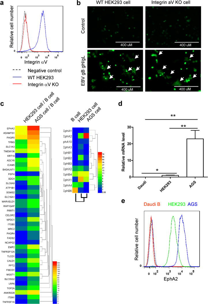
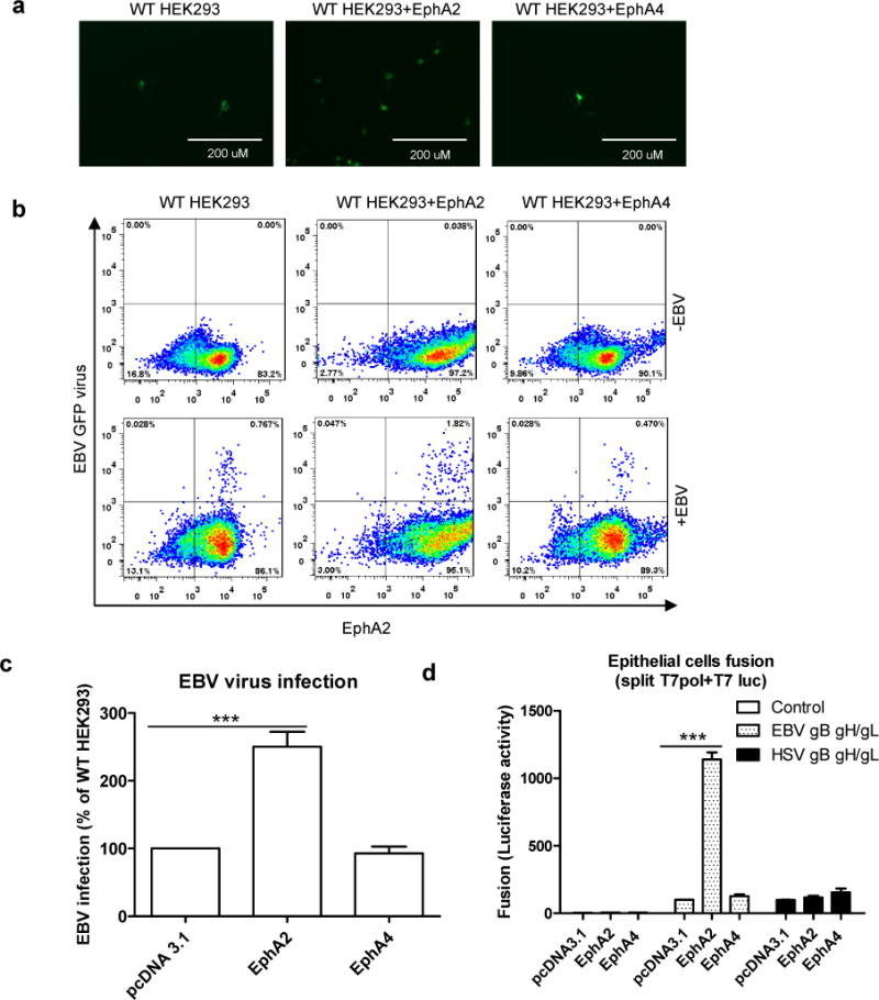
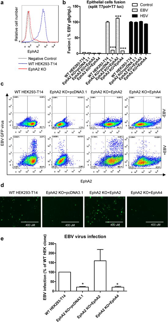
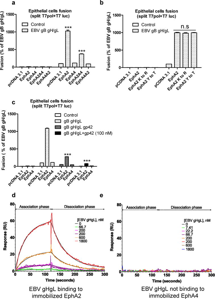
Epstein-Barr virus (EBV) is an oncogenic virus that infects more than 90% of the world's population 1 . EBV predominantly infects human B cells and epithelial cells, which is initiated by fusion of the viral envelope with a host cellular membrane 2 . The mechanism of EBV entry into B cells has been well characterized 3 . However, the mechanism for epithelial cell entry remains elusive. Here, we show that the integrins αvβ5, αvβ6 and αvβ8 do not function as entry and fusion receptors for epithelial cells, whereas Ephrin receptor tyrosine kinase A2 (EphA2) functions well for both. EphA2 overexpression significantly increased EBV infection of HEK293 cells. Using a virus-free cell-cell fusion assay, we found that EphA2 dramatically promoted EBV but not herpes simplex virus (HSV) fusion with HEK293 cells. EphA2 silencing using small hairpin RNA (shRNA) or knockout by CRISPR-Cas9 blocked fusion with epithelial cells. This inhibitory effect was rescued by the expression of EphA2. Antibody against EphA2 blocked epithelial cell infection. Using label-free surface plasmon resonance binding studies, we confirmed that EphA2 but not EphA4 specifically bound to EBV gHgL and this interaction is through the EphA2 extracellular domain (ECD). The discovery of EphA2 as an EBV epithelial cell receptor has important implications for EBV pathogenesis and may uncover new potential targets that can be used for the development of novel intervention strategies.


Conflict of interest statement

Competing interests

The authors declare no competing financial interests.

Figures

Figure 1
Figure 1. Identification of EphA2 as the potential EBV epithelial cell receptor
a, Integrin aV cell surface expression in integrin aV WT and KO HEK293 cells by flow cytometry. b, CHO-K1 cells transfected with GFP plasmid plus either control plasmid or EBV gHgL and gB were overlaid with integrin aV WT and KO HEK293 cells. Syncytia formation was visualized and captured with a EVOS fluorescence microscope (representative data from three independent experiments). c, Heat map of the normalized expression of potential epithelial cell receptors compared to B cells for HEK293 and AGS cells (left). Heat map of Ephs for B cells, HEK293 cells, and AGS cells (right). d, Comparison of EphA2 expression in different cell types. Total RNA from each cell type was assayed for EphA2 and GAPDH mRNA levels by quantitative RT-PCR. The bars represent the relative EphA2 mRNA expression that was normalized to GAPDH. Data are means plus standard errors of the means for three independent experiments. e, Flow cytometry histograms of EphA2 in Daudi B, HEK293 and AGS cells, with the dotted line as a negative control. One representative flow cytometry data of at least two independent experiments is shown in a and e. *P<0.05 and **P<0.01 (ANOVA followed by post-hoc Tukey’s multiple comparison test).
Figure 2
Figure 2. EphA2 can promote both EBV infection and virus-free cell-cell fusion
a, HEK293 cells were transfected with pcDNA 3.1, EphA2 or EphA4. 24h post transfection, 5×10⁴ cells were seeded onto a 48 well plate. 24 hours later, the cells were infected with 100 mL EBfaV-GFP virus concentrated from 1 mL virus-containing supernatant. 72 hours later, the infected GFP cells were visualized and captured with a EVOS fluorescence microscope (a) or analyzed by flow cytometry (b). Data are representative data from three independent experiments. c, Quantification of the flow cytomery data from three independent experiments, the bars represent the percentage of infection with infection of pcDNA 3.1 transfected HEK293 cells set to 100%. d, CHO-K1 cells transfected with T7 luciferase plasmid together with either control plasmid, EBV gHgL and gB, or HSV gHgL, gB and gD were overlaid with HEK293 cells transfected with pcDNA 3.1, EphA2 or EphA4. EBV fusion with HEK293 cells transfected with pcDNA 3.1 was set to 100%. HSV fusion was normalized to EBV fusion and standardized with pcDNA 3.1 transfected cells set to 100%. Data are means plus standard errors of the means for three independent experiments for c and d. ***P<0.001 (ANOVA followed by post-hoc Tukey’s multiple comparison test).
Figure 3
Figure 3. EphA2 is essential for EBV infection and virus-free cell-cell fusion
a, EphA2 cell surface expression in EphA2 WT and KO HEK293-T14 cells by flow cytometry. One representative flow cytometry data of three independent experiments is shown. b, Virus-free EBV or HSV fusion with EphA2 WT and KO HEK293-T14 cells or EphA2 KO cells that overexpress EphA2 or EphA4. The bars represent the fusion activity and data are means plus standard errors of the means for three independent experiments. c-e, EphA2 WT and KO HEK293-T14 cells or EphA2 KO cells that overexpress EphA2 or EphA4 were infected with EBfaV-GFP virus. 72 h post infection, the infected GFP cells were visualized and captured with a EVOS fluorescence microscope (d) or GFP-positive cells were analyzed by flow cytometry (c). Data are representative data from three independent experiments. e, Quantification of the flow cytometry data from three independent experiments, the bars represent the percentage of infection and infection of WT HEK293-T14 cells was set to 100. Data are means plus standard errors of the means. *P<0.05 and ***P<0.001 vs WT HEK293-T14 (ANOVA followed by post-hoc Tukey’s multiple comparison test).
Figure 4
Figure 4. EphA2 binds to EBV gHgL with low affinity
a, virus-free EBV fusion with HEK293 cells transfected with pcDNA 3.1, EphA2, EphA4 or EphA2 and EphA4 chimeras as indicated. b, virus-free EBV fusion with HEK293 cells transfected with control, EphA2, EphA4 or EphA2 kinase-dead mutants as indicated. Fusion activity of HEK293 cells transfected with pcDNA 3.1 was set to 100 (a) and (b). c, HEK293 cells transfected with T7 polymerase together with pcDNA 3.1, EphA2, or EphA4 were overlaid with CHO-K1 cells transfected with T7 luciferase plasmid together with EBV gB, gHgL in the presence or absence of gp42 plasmid or 100 nM soluble gp42. EBV fusion with HEK293 cells transfected with pcDNA 3.1 was set to 100. The bars represent the fusion activity (a, b and c) and data are means plus standard errors of the means for three independent experiments. d and e, EphA2 and EphA4 were immobilized on two separate channels of a CMD biosensor chip surface. Label-free SPR binding kinetics of EBV gHgL with EphA2 (d) or EphA4 (e) are depicted with model fits represented by dashed line. Global curve fitting with a 1:1 interaction model (dashed lines) closely matches the experimental data for EphA2 binding to EBV gHgL. EphA4 does not bind to EBV gHgL under identical conditions (representative data from three independent experiments). ***P<0.001 vs EBV gHgL, pcDNA 3.1 in a, vs gB gHgL, EphA2 in c (ANOVA followed by post-hoc Tukey’s multiple comparison test).

References

    1. Longnecker R, Kieff E, Cohen JI. Fields Viorology. 6. Lippincott Williams & Wilkins; Philadelphia, PA: 2013.
    1. Connolly SA, Jackson JO, Jardetzky TS, Longnecker R. Fusing structure and function: a structural view of the herpesvirus entry machinery. Nature reviews Microbiology. 2011;9(5):369–381. - PMC - PubMed
    1. Sathiyamoorthy K, Jiang J, Hu YX, Rowe CL, Möhl BS, Chen J, et al. Assembly and architecture of the EBV B cell entry triggering complex. 2014 - PMC - PubMed
    1. Epstein MA, Achong BG, Barr YM. Virus Particles in Cultured Lymphoblasts from Burkitt’s Lymphoma. Lancet. 1964;1(7335):702–703. - PubMed
    1. Fingeroth JD, Weis JJ, Tedder TF, Strominger JL, Biro PA, Fearon DT. Epstein-Barr virus receptor of human B lymphocytes is the C3d receptor CR2. Proceedings of the National Academy of Sciences of the United States of America. 1984;81(14):4510–4514. - PMC - PubMed

Publication types

MeSH terms