. 2018 Feb 14;118(3):919-988.
doi: 10.1021/acs.chemrev.6b00750. Epub 2018 Jan 2.

Protein Lipidation: Occurrence, Mechanisms, Biological Functions, and Enabling Technologies

Affiliations

Protein Lipidation: Occurrence, Mechanisms, Biological Functions, and Enabling Technologies

Hong Jiang et al. Chem Rev. .

Abstract

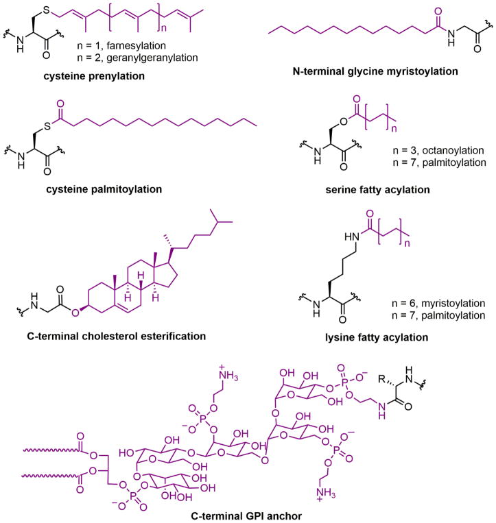
Protein lipidation, including cysteine prenylation, N-terminal glycine myristoylation, cysteine palmitoylation, and serine and lysine fatty acylation, occurs in many proteins in eukaryotic cells and regulates numerous biological pathways, such as membrane trafficking, protein secretion, signal transduction, and apoptosis. We provide a comprehensive review of protein lipidation, including descriptions of proteins known to be modified and the functions of the modifications, the enzymes that control them, and the tools and technologies developed to study them. We also highlight key questions about protein lipidation that remain to be answered, the challenges associated with answering such questions, and possible solutions to overcome these challenges.


Figures

Figure 1
Figure 1
Lipid modifications of proteins. GPI, glycosylphosphatidylinositol.
Figure 2
Figure 2
Protein prenylation.
Figure 3
Figure 3
(A) Protein structures of FT (PDB ID 1FT1), GTT-1 (PDB ID 1N4P), and RGGT (PDB ID 1DCE). The α subunits (green) of FT and GTT-1 are identical. There are extra leucine-rich repeats (LRRs) and immunoglobulin (Ig)-like domains in the α subunit of RGGT (α-helices in cyan and β-sheets in red). (B) Superimposition of the β subunits of FT (cyan), GGT-1 (yellow), and RGGT (magenta) to show the structural homology. (C) The binding of substrates versus product in GGT-1. Geranylgeranyl diphosphate (GGPP; indicated by a GGPP analogue in magenta) rotates toward the cysteine in the CaaX peptide (PDB ID 1N4Q) to form the prenylated product (green; PDB ID 1N4R). (D) Simultaneous binding of GGPP (magenta) and the translocated prenylated product (green) at the active site of GGT-1 (PDB ID 1N4S). (E) The zinc binding site in the β subunit of FT (PDB ID 1D8D). (F) In FT, GGT-1, and RGGT, conserved residues in the β subunits of prenyltransferases bind to zinc, including an aspartate residue (Asp297β, Asp269β, and Asp238β, respectively), a cysteine residue (Cys299β, Cys271β, and Cys240β, respectively), and a histidine residue (His362β, His321β, and His290β, respectively). The zinc also coordinates with the cysteine residue of CaaX peptides. (G) Binding position of isoprenoid diphosphate in prenyltransferases, including FPP in FT (PDB ID 1FT2) and GGPP in GGT-1 (PDB ID 1N4P) and RGGT (PDB ID 3DST). (H) Comparison of isoprenoid diphosphate binding in FT (PDB ID 1FT2), GGT-I (PDB ID 1N4P), and RGGT (PDB ID 3DST). FPP (pink) with Trp102β and Tyr361β (pink) in FT, GGPP (green) with Thr49β and Phe324β (green) in GGT-1, and GGPP (yellow) with Ser48β and Phe293β (yellow) in RGGT. In FT, the bulky Trp102β residue occupies the space in which the fourth isoprene unit of GGPP binds in GGT-1 and RGGT. This residue determines the isoprenoid specificity. (I) Protein structure of the RGGT-REP-1 complex (PDB ID 1LTX). REP-1 is yellow. (J) Protein structure of the prenylated Rab7-REP-1 complex (PDB ID 1VG0). REP-1 is yellow and Rab7 is blue. All protein structures were made using PyMol with the PDB files.
Figure 4
Figure 4
General reaction scheme with an ordered sequential kinetic mechanism for prenylation catalyzed by FT and GGT-1. The kinetics data for farnesylation and geranylgeranylation are from reference and , respectively.
Figure 5
Figure 5
Reaction pathway of Rab digeranylgeranylation catalyzed by RGGT.
Figure 6
Figure 6
Chemical probes used to study protein prenylation.
Figure 7
Figure 7
Plasma membrane targeting involving prenylation and a second signal, including (I) upstream palmitoylation, (II) downstream palmitoylation, and (III) upstream polybasic domain (typically six lysine residues).
Figure 8
Figure 8
Protein structures of guanosine diphosphate dissociation inhibitors (GDIs) in complex with prenylated proteins. (A) Prenylated Cdc42 (green)-RhoGDI (cyan) complex (PDB ID 1DOA), (B) prenylated Rac1 (green)–RhoGDI (cyan) complex (PDB ID 1HH4), (C) prenylated RhoA (green)-RhoGDI (cyan) complex (PDB ID 4F38), (D) prenylated Rheb (green)-PDEδ (cyan) complex (PDB ID 3T5G), (E) prenylated YPT1 (green)-RabGDI (cyan) complex (PDB ID 1UKV), and (F) doubly prenylated YPT1 (green)-RabGDI (cyan) complex (PDB ID 2BCG). CBR, C-terminal-binding region. The prenyl moiety is shown in purple or red. All protein structures were made using PyMOL with PDB files.
Figure 9
Figure 9
Mechanism of RhoA membrane extraction by RhoGDI. GG, geranylgeranyl group.
Figure 10
Figure 10
Farnesyltransferase inhibitors.
Figure 11
Figure 11
Specific inhibitors of GGT-1 and RGGT and dual inhibitors of FT and GGT-1. IC50, half-maximal inhibitory concentration.
Figure 12
Figure 12
(A) Myristoyl modification at an N-terminal glycine residue. (B) Co-translational N-myristoyl modification. (C) Post-translational N-myristoyl modification.
Figure 13
Figure 13
(A) Crystal structure of S. cerevisiae NMT in complex with a non-hydrolyzable myristoyl-CoA analogue and a peptide substrate (PDB ID 1IID). (B) Phe170 and Leu171 form the oxyanion hole to stabilize the negative charge developed on the carbonyl oxygen of myristoyl-CoA during catalysis. (C) The hydrophobic myristoyl group binds in a deep pocket in NMT. (D) The peptide substrate recognition site of NMT, which explains the peptide sequence specificity of NMT. All protein structures were made using PyMOL with PDB files.
Figure 14
Figure 14
Myristoyl switch mechanisms. (A) The phosphorylation of N-glycine myristoylated protein stimulates membrane dissociation by interrupting the electrostatic interaction between proteins and the phospholipid. (B) Ligand binding enhances the membrane association of N-glycine myristoylated proteins. (C) Proteolysis triggers the release of N-glycine myristoylated protein from the membrane.
Figure 15
Figure 15
N-Glycine myristoylation may facilitate the trans interaction between Golgi reassembly stacking proteins by limiting conformational flexibility.
Figure 16
Figure 16
The myristoyl switch that regulates c-Abl activity. The c-Abl structure (PDB ID 1OPL) in complex with myristoyl and a kinase inhibitor is superimposed on the c-Abl structure without bound myristoyl (PDB ID 1M52). In the absence of myristoyl, an extended α-helix (αI, grey) prevents the binding of the SH2 domain to the kinase domain. In the myristoyl-bound state, the αI helix is separated into two shorter helices, αI (magenta) and αI′ (blue). The αI′ helix makes an abrupt turn to bind to the myristoyl group. This conformational change leads to the docking of the SH2 domain at the kinase domain and subsequent autoinhibition.
Figure 17
Figure 17
Structures and half-maximal inhibitory concentration (IC50) values of representative inhibitors developed for NMTs in various species (CaNMT: Candida albicans NMT; HsNMT1/2, Homo sapiens NMT1/2; PfNMT, Plasmodium falciparum NMT; PvNMT, Plasmodium vivax NMT; and TbNMT: Trypanosoma brucei NMT).
Figure 18
Figure 18
Reversible cysteine palmitoylation.
Figure 19
Figure 19
Predicted topology and domain structure of DHHCs. TMD, transmembrane domain.
Figure 20
Figure 20
Mechanism of DHHC-catalyzed cysteine palmitoylation.
Figure 21
Figure 21
Inhibitors for cysteine depalmitoylases APT1, APT2, and ABHD17.
Figure 22
Figure 22
Structures of reported palmitoylation inhibitors.
Figure 23
Figure 23
C-terminal sequences of Ras family members and Ras trafficking.
Figure 24
Figure 24
Bioorthogonal palmitic acid probes for the detection of protein palmitoylation.
Figure 25
Figure 25
Procedure of ABE method for the detection of protein S-palmitoylation.
Figure 26
Figure 26
Method for imaging palmitoylated proteins in cells.
Figure 27
Figure 27
Protein O- and N-acylation and protein C-terminal cholesterol esterification.
Figure 28
Figure 28
Inhibitors targeting Porcupine (PORCN) and Hedgehog acyltransferase (Hhat).
Figure 29
Figure 29
Crystal structure of Xenopus Wnt8 in complex with the Frizzled-8 (Fz8) cysteine-rich domain (CRD; PDB 4F0A). CTD, C-terminal domain; NTD, N-terminal domain.
Figure 30
Figure 30
Two proposed mechanism for the N-palmitoylation of Hedgehog (Hh) proteins.
Figure 31
Figure 31
Mechanism of C-terminal autoprocessing of Hh proteins.

References

    1. Lemmon MA, Ferguson KM. Signal-Dependent Membrane Targeting by Pleckstrin Homology (Ph) Domains. Biochem J. 2000;350(Pt 1):1–18. - PMC - PubMed
    1. Lentz BR. Exposure of Platelet Membrane Phosphatidylserine Regulates Blood Coagulation. Prog Lipid Res. 2003;42:423–438. - PubMed
    1. Menon AK, Kinoshita T, Orlean PA, Tamanoi F. Glycosylphosphatidylinositol (Gpi) Anchoring of Proteins. Academic Press; 2009.
    1. Zurzolo C, Simons K. Glycosylphosphatidylinositol-Anchored Proteins: Membrane Organization and Transport. Biochim Biophys Acta. 2016;1858:632–639. - PubMed
    1. Paladino S, Lebreton S, Zurzolo C. Trafficking and Membrane Organization of Gpi-Anchored Proteins in Health and Diseases. Curr Top Membr. 2015;75:269–303. - PubMed

Publication types