Review
. 2018 Jan 2;128(1):64-73.
doi: 10.1172/JCI93560. Epub 2018 Jan 2.

Endoplasmic reticulum stress in the pathogenesis of fibrotic disease

Affiliations
Review

Endoplasmic reticulum stress in the pathogenesis of fibrotic disease

Jonathan A Kropski et al. J Clin Invest. .

Abstract

Eukaryotic cells contain an elegant protein quality control system that is crucial in maintaining cellular homeostasis; however, dysfunction of this system results in endoplasmic reticulum (ER) stress and activation of the unfolded protein response (UPR). Severe or prolonged ER stress is associated with the development of degenerative and fibrotic disorders in multiple organs, as evidenced by the identification of disease-causing mutations in epithelial-restricted genes that lead to protein misfolding or mistrafficking in familial fibrotic diseases. Emerging evidence implicates ER stress and UPR signaling in a variety of profibrotic mechanisms in individual cell types. In epithelial cells, ER stress can induce apoptosis, inflammatory signaling, and epithelial-mesenchymal transition. In other cell types, ER stress is linked to myofibroblast activation, macrophage polarization, and T cell differentiation. ER stress-targeted therapies have begun to emerge using approaches that range from global enhancement of chaperone function to selective targeting of activated ER stress sensors and other downstream mediators. As the complex regulatory mechanisms of this system are further clarified, there are opportunities to develop new disease-modifying therapeutic strategies in a wide range of chronic fibrotic diseases.


Conflict of interest statement

Conflict of interest: The authors have declared that no conflict of interest exists.

Figures

Figure 1
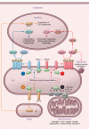
Figure 1. Overview of ER stress–related signaling.
Bip binds to accumulating misfolded proteins in the ER, leading to its dissociation from the three ER stress sensors, IRE1α, PERK, and ATF6. (i) Dissociation from Bip allows IRE1α to multimerize and autophosphorylate, activating endoribonuclease activity that leads to alternative splicing of the transcription factor XBP1. Spliced XBP1 (XBP1s) then translocates to the nucleus and promotes transcription of components of the ERAD system. Oligomerized IRE1α loses stringency of endoribonuclease activity and activates regulated IRE1-dependent decay (RIDD), thereby degrading mRNA and miRNAs. (ii) Bip dissociation leads to dimerization and autophosphorylation of PERK, which phosphorylates eIF2α to inhibit protein translation and signals for ATF4 nuclear translocation. Once in the nucleus, ATF4 activates ATF3, which induces adaptive antioxidant responses, promotes amino acid synthesis, and promotes autophagy. (iii) Bip dissociation from ATF6 permits its transit from the ER to the Golgi, where further processing allows trafficking to the nucleus and subsequent increases in production of ER chaperones. (iv) Bip and other ER chaperones serve as calcium-binding proteins. The ER tightly controls the cytosolic calcium pool available for mitochondrial uptake through the mitochondrial calcium uniporter (MCU) via sarcoendoplasmic reticulum Ca2+ ATPase (SERCA) and the inositol triphosphate receptor (IP3R). Through its regulation of calcium flux, the ER plays a central role in the regulation of cellular bioenergetics and mitochondrial mechanisms of apoptosis.
Figure 2
Figure 2. Mechanisms of fibrosis related to ER stress.
In epithelial cells, ER stress induces a profibrotic microenvironment by promoting apoptosis, suppressing progenitor cell function, activating inflammatory signaling pathways, and inducing production of profibrotic mediators that promote fibroblast proliferation and myofibroblast differentiation. ER stress signaling in T lymphocytes suppresses Th1 and Th2 polarization and drives Th17 polarization, which can promote fibrosis through interactions with epithelium and fibroblasts. In macrophages, ER stress facilitates acquisition of the M2 phenotype, which is accompanied by enhanced production of profibrotic mediators.

References

    1. Rockey DC, Bell PD, Hill JA. Fibrosis--a common pathway to organ injury and failure. N Engl J Med. 2015;372(12):1138–49. doi: 10.1056/NEJMra1300575. - DOI - PubMed
    1. Wynn TA. Common and unique mechanisms regulate fibrosis in various fibroproliferative diseases. J Clin Invest. 2007;117(3):524–9. doi: 10.1172/JCI31487. - DOI - PMC - PubMed
    1. Nanthakumar CB, Hatley RJD, Lemma S, Gauldie J, Marshall RP, Macdonald SJF. Dissecting fibrosis: therapeutic insights from the small-molecule toolbox. Nat Rev Drug Discov. 2015;14(10):693–720. doi: 10.1038/nrd4592. - DOI - PubMed
    1. López-Otín C, Blasco MA, Partridge L, Serrano M, Kroemer G. The hallmarks of aging. Cell. 2013;153(6):1194–217. doi: 10.1016/j.cell.2013.05.039. - DOI - PMC - PubMed
    1. Kropski JA, Lawson WE, Young LR, Blackwell TS. Genetic studies provide clues on the pathogenesis of idiopathic pulmonary fibrosis. Dis Model Mech. 2013;6(1):9–17. doi: 10.1242/dmm.010736. - DOI - PMC - PubMed

Publication types

Substances