Review
. 2018 Jan 2;128(1):54-63.
doi: 10.1172/JCI93558. Epub 2018 Jan 2.

The perivascular origin of pathological fibroblasts

Review

The perivascular origin of pathological fibroblasts

Selene E Di Carlo et al. J Clin Invest. .

Abstract

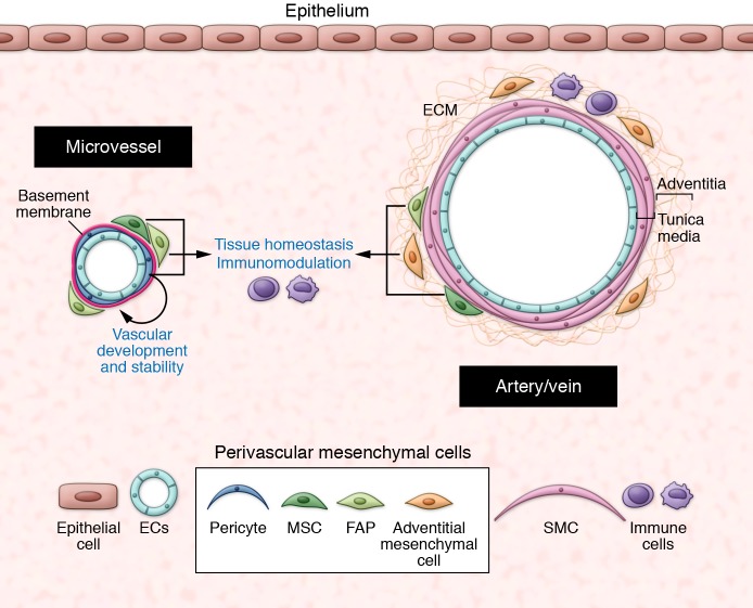
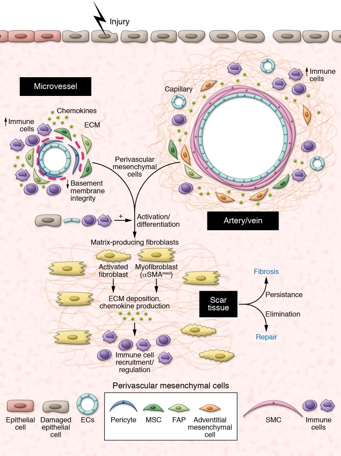
The ability to repair tissues is essential for the survival of organisms. In chronic settings, the failure of the repair process to terminate results in overproduction of collagen, a pathology known as fibrosis, which compromises organ recovery and impairs function. The origin of the collagen-overproducing cell has been debated for years. Here we review recent insights gained from the use of lineage tracing approaches in several organs. The resulting evidence points toward specific subsets of tissue-resident mesenchymal cells, mainly localized in a perivascular position, as the major source for collagen-producing cells after injury. We discuss these findings in view of the functional heterogeneity of mesenchymal cells of the perivascular niche, which have essential vascular, immune, and regenerative functions that need to be preserved for efficient repair.


Conflict of interest statement

Conflict of interest: L. Peduto is co-inventor of the patent (no. 9777276) “The use of ADAM12 inhibitors to control inflammation-induced fibrosis” at Institut Pasteur.

Figures

Figure 1
Figure 1. The perivascular niche.
At steady state, the perivascular niche contains different subsets of mesenchymal cells, depending on the vessel type and size. In the microvasculature, pericytes are embedded within the vascular basement membrane and are essential for vascular development and stability. The outer covering of larger vessels (the adventitia) contains adventitial mesenchymal cells in a collagenous matrix. Other mesenchymal subsets such as FAPs and MSCs are localized in close proximity to blood vessels. At steady state, perivascular mesenchymal cells have essential roles in maintenance of tissue homeostasis. SMC: smooth muscle cells.
Figure 2
Figure 2. The perivascular niche after injury.
Injury induces activation and differentiation of specific subsets of perivascular mesenchymal cells toward a myofibroblastic phenotype, which regulate scar tissue formation and immune cells recruitment/activity through production of ECM, chemokines and growth factors. Whether all perivascular mesenchymal cell types are similarly involved in this process remain unclear. Signals produced by damaged epithelial cells, endothelial cells, and inflammatory cells further contribute to this transition. Failure to terminate this repair program leads to fibrosis.

References

    1. Hinz B, Phan SH, Thannickal VJ, Galli A, Bochaton-Piallat ML, Gabbiani G. The myofibroblast: one function, multiple origins. Am J Pathol. 2007;170(6):1807–1816. doi: 10.2353/ajpath.2007.070112. - DOI - PMC - PubMed
    1. Hinz B, et al. Recent developments in myofibroblast biology: paradigms for connective tissue remodeling. Am J Pathol. 2012;180(4):1340–1355. doi: 10.1016/j.ajpath.2012.02.004. - DOI - PMC - PubMed
    1. Darby I, Skalli O, Gabbiani G. α-Smooth muscle actin is transiently expressed by myofibroblasts during experimental wound healing. Lab Invest. 1990;63(1):21–29. - PubMed
    1. Hinz B. Formation and function of the myofibroblast during tissue repair. J Invest Dermatol. 2007;127(3):526–537. doi: 10.1038/sj.jid.5700613. - DOI - PubMed
    1. Sun KH, Chang Y, Reed NI, Sheppard D. α-Smooth muscle actin is an inconsistent marker of fibroblasts responsible for force-dependent TGF-β activation or collagen production across multiple models of organ fibrosis. Am J Physiol Lung Cell Mol Physiol. 2016;310(9):L824–L836. doi: 10.1152/ajplung.00350.2015. - DOI - PMC - PubMed

Publication types